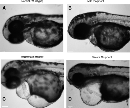
FIG. 8.
Heart defects observed in morphants following rescue experiments with Myh6 mRNA in zebrafish. (A) Normal phenotype. Both the atrium and the ventricle are clearly defined (in some cases, it is even possible to observe myocardial and endocardial layers) and tightly packed into a figure eight configuration. (B) Mild phenotype. The atrium and ventricle are clearly distinguishable, but looping is noticeably relaxed and slight pericardial edema is seen. (C) Moderate phenotype. The atrium and ventricle can still be identified but are irregular in shape and/or size. Looping is lost, and pericardial edema is observed. (D) Severe phenotype. Complete malformation is seen, with no defined atrium or ventricle. The heart is stretched to a thin tube. Large pericardial edema is often observed, and the heart rate is notably slower. Many other morphological observations may also be seen (e.g., spinal defects).

