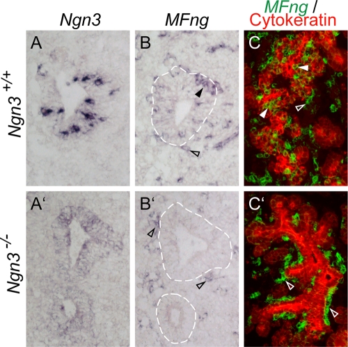
FIG. 2.
Loss of MFng expression in Ngn3−/− pancreas. E10.5 (A to B′) and E14.5 (C and C′) pancreases of wild-type (A to C) and Ngn3-deficient (A′ to C′) mice were analyzed by in situ hybridization. (A and B) In wild-type pancreas at E10.5, Ngn3 and MFng mRNAs (blue) are detected in scattered epithelial cells. The expression of MFng is also detected in mesenchymal cells (open arrowhead in panel B). (A′ and B′) In Ngn3−/− pancreatic epithelia at E10.5, mutant Ngn3 mRNA is weakly detected (blue in panel A′) whereas MFng mRNA (blue in panel B′) is not detected. The expression of MFng (open arrowheads in panel B′) is retained in mesenchymal cells. (C and C′) In wild-type E14.5 embryos, MFng (green pseudocolor) is expressed both in the pancytokeratin-positive pancreatic epithelium (red) and within the surrounding mesenchyme (C). In Ngn3−/− pancreas, MFng expression is retained only in the mesenchymal cells (C′). Broken lines in panels B and B′ delineate pancreatic epithelia. Closed arrowheads mark MFng-positive pancreatic epithelial tissue. Open arrowheads mark MFng-positive pancreatic mesenchyme tissue.

