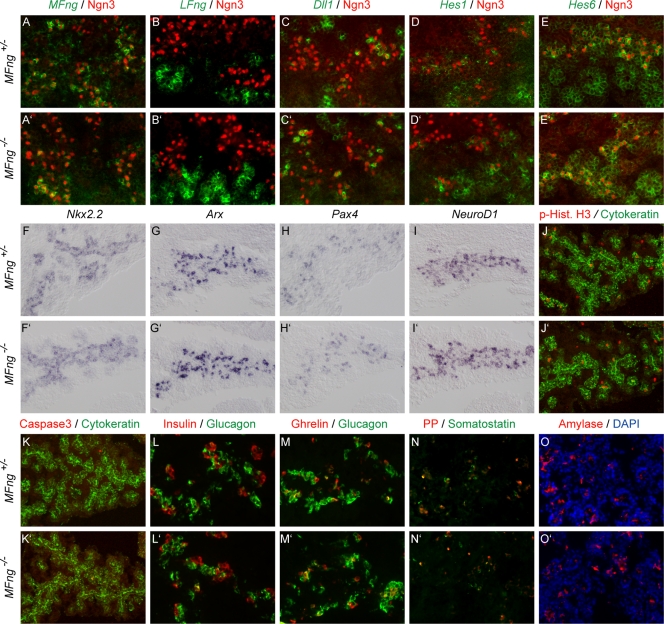
FIG. 6.
Normal pancreatic development in MFng−/− mice. (A to E′) In situ hybridization of Notch signaling genes (green pseudocolor in A to E′) in E14.5 pancreases and proendocrine cells expressing Ngn3 (red immunohistochemistry staining) in MFng+/− (A to E) and MFng−/− (A′ to E′) mice. Mutant MFng mRNA was detected at reduced levels (compare panels A and A′), no compensatory upregulation of LFng (green pseudocolor in panels B and B′) was observed, and the expression of Dll1 and Hes1 and Hes6 (as indicated in panels C to E′) was unaltered. (F to I′) In situ hybridization of progenitor cell markers Nkx2.2, Arx, Pax4, and NeuroD1 (blue, as indicated). (J to K′) Immunohistochemical analysis of proliferation and apoptosis markers phospho-histone H3 (red in panels J and J′) and cleaved caspase 3 (red in panels K and K′) in E14.5 pancreatic epithelia (indicated by green pancytokeratin immunohistochemistry staining). (L to O′) Immunohistochemical analyses of endocrine hormones (as indicated in panels L to N′) and the exocrine enzyme amylase (O and O′). Nuclear staining with DAPI (blue) is shown in panels O and O′.

