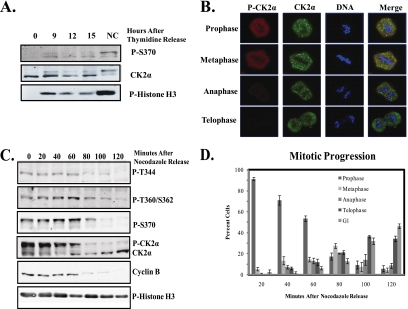
FIG. 2.
CK2α phosphorylation occurs in mitotic cells during prophase and metaphase. (A) U2OS cells were synchronized in S phase by a double-thymidine block and released into mitosis for 9, 12, or 15 h before harvest. Lysates were immunoblotted with phosphospecific CK2α antibody P-S370. Total CK2α is also shown. Mitotic cells were detected with a phospho-histone H3 (Ser 10) antibody. (B) U2OS cells were synchronized as in panel A, fixed 12 h after thymidine release, and immunostained with antibodies against phosphorylated CK2α (P-CK2α) (red) and total CK2α (green). DNA was stained with DAPI. Magnification, ×63. (C) U2OS cells were arrested in mitosis by nocodazole treatment, washed, and harvested at 20-min intervals after the removal of nocodazole. Lysates were immunoblotted with antibodies against phosphorylated CK2α, total CK2α, cyclin B1 to detect the onset of anaphase, and phospho-histone H3 (serine 10) to detect mitotic cells. (D) Cells were arrested in mitosis as in panel C and plated on slides following release. After fixation and DAPI staining, cells were scored for mitotic stage based on DNA morphology. At least 100 cells were scored for each time point in each of three replicate experiments.

