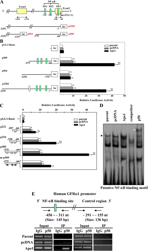
FIG. 2.
Ape1/Ref-1 increases GFRα1 promoter activity by enhancing p50 NF-κB activation. (A) The upper panel shows a schematic representation of the human GFRα1 promoter region. The three putative NF-κB-binding sites spanning positions −349 to −335 (NF1), −300 to −287 (NF2), and −155 to −143 (NF3) are shown in green. The middle and lower panels show the GFRα1 promoter region of the promoter-reporter constructs p2284 (positions −2291 to −7), p964 (positions −2291 to −1327), and p509 (positions −575 to −66). (B) Parent cells and pcDNA3- and Ape1/Ref-11-expressing cells were transfected with pGL3-Basic, p2284, p964, or p509. Representations of the promoters are shown. The values reported are means ± the SD from six separate experiments. **, P < 0.01. (C) Cells were transfected with pGL3-Basic, p509 (positions −575 to −66), p221 (positions −287 to −66 of p509), p254 (positions −320 to −66 of p509), or m-p509 (p509 containing three mutated NF-κB-binding motifs). Schematic representations of the promoters are shown; ovals represent the consensus sites for the NF-κB-binding motifs. The values shown are means ± the SD from six separate experiments. **, P < 0.01. (D) Biotin-labeled probes oligonucleotides containing the putative NF-κB-binding site (NF1) from the GFRα1 promoter region were incubated with nuclear extracts prepared from parent cells and pcDNA3- and Ape1/Ref-1-transfected cells. Unlabeled oligonucleotides were used as a competitor. For supershift assays, anti-p50 or anti-p52 antibodies were added to the reaction mixtures, followed by incubation for 30 min prior to separation of the DNA-protein complexes. Black and gray triangles indicate DNA-protein complexes and antibody-supershifted complexes, respectively. (E) At the top of the panel is a schematic representation of the human GFRα1 promoter region. Chromatin from parent cells and pcDNA3- and Ape1/Ref-1-expressing GM00637 cells were fixed with formaldehyde and fragmented by sonication. The fragmented chromatin was immunoprecipitated using anti-p50 antibody and anti-rabbit IgG and then analyzed by PCR using primer sets specific for the promoter region containing putative NF-κB-binding site (green box: NF1) or control region, as indicated by the arrow.

