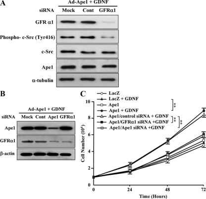
FIG. 3.
Ape1/Ref-1 increases c-Src phosphorylation and cellular proliferation in response to GDNF through GFRα1. (A) Ad-Ape1/Ref-1-infected GM00637 cells were transfected with the mock, control siRNA (Cont), or GFRα1 siRNA. At 48 h after transfection, cells were incubated with GDNF (10 ng/ml) for 1 h. Equal amounts (20 μg of proteins) of the cell lysates were separated by SDS-10% PAGE and then transferred onto a nitrocellulose membrane. The membrane was immunoblotted with anti-Ape1/Ref-1, anti-GFRα1, or anti-phospho c-Src (Tyr418) antibodies. To control for equal loading, the membrane was stripped and reprobed against nonphosphorylated c-Src and α-tubulin. (B) Ad-Ape1/Ref-1-infected GM00637 cells were transfected with the mock, control-siRNA (Cont), Ape1/Ref-1-siRNA (Ape1), or GFRα1-siRNA. At 48 h after transfection, cells were then incubated with GDNF (10 ng/ml) for an additional 24 h. The total protein was extracted from the cells, and equal amounts (20 μg of proteins) of the cell lysates were separated by SDS-10% PAGE and then transferred onto a nitrocellulose membrane. The membrane was immunoblotted with anti-Ape1/Ref-1, anti-GFRα1, or anti-β-actin antibodies. (C) Ad-LacZ (LacZ)- or Ad-Ape1/Ref-1 (Ape1)-infected GM00637 cells were transfected with control siRNA or GFRα1 siRNA and then incubated with or without GDNF (10 ng/ml) for up to 72 h. The number of cells was determined by counting the cells every 24 h after GDNF treatment. Each value is the mean ± the SD from three separate experiments. **, P < 0.01.

