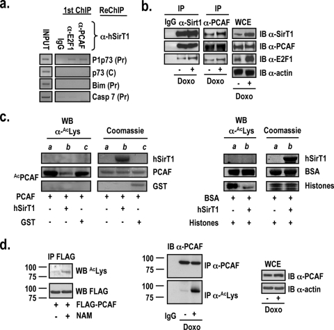
FIG. 5.
hSirT1, PCAF, and E2F1 interact and are corecruited onto the P1p73 promoter. (a) Chromatin from asynchronously growing U2Os cells was analyzed by sequential ChIP using either anti-E2F1 or anti-PCAF (first immunoprecipitation) and anti-hSirT1 (second immunoprecipitation). IgG, control immunoprecipitation with the relevant control IgGs. ChIP DNA was amplified with primers specific for the P1p73, Bim, and caspase 7 promoter regions containing E2F1 sites (Pr) and with the corresponding control distant oligonucleotides (C) (data not shown). (b) Extracts from asynchronously growing or doxorubicin (Doxo) (2 μM)-treated U2Os cells were immunoprecipitated (IP) with control IgG, anti-hSirT1, and anti-PCAF antibodies and immunoblotted (IB) with anti-hSirT1 (α-SirT1), anti-PCAF, and anti-E2F1 antibodies. (c) hSirT1 deacetylates PCAF in vitro. (Left) Baculovirus affinity-purified full-length FLAG-PCAF (kindly provided by V. Sartorelli, NIAMS, NIH) and purified active bacterial GST and GST-tagged human SirT1 (amino acids 193 to 741) were employed in the acetylation (lane a) and deacetylation (lanes b and c) reactions in vitro. Reaction products were separated by SDS-polyacrylamide gel electrophoresis and immunoblotted with the anti-acetyl-Lys antibody. Lane a shows in vitro PCAF autoacetylation. (Right) Acetylated BSA and acetylated histones (lane a) were incubated with GST-tagged human SirT1 (amino acids 193 to 741) (Upstate Inc.) in the deacetylation reaction (see b). Coomassie-stained gels show the input proteins used in the reactions. WB, Western blot. (d) hSirT1 regulates PCAF acetylation in vivo. (Left) 293 cells were transfected with the FLAG-PCAF vector and exposed to NAM (10 mM). Extracts were immunoprecipitated with the M2 FLAG antibody and immunoblotted with anti-acetyl-Lys antibody. (Middle) PCAF acetylation increases in response to DNA damage. Extracts from untreated and doxorubicin (2 μM)-treated U2Os cells were immunoprecipitated with either anti-PCAF or anti-acetyl-Lys antibody and immunoblotted with the anti-PCAF antibody. (Right) Total PCAF protein levels were analyzed by immunoblotting in whole-cell extracts (WCE) from untreated and doxorubicin-treated cells. Molecular weight positions (in thousands) in the SDS-polyacrylamide gels are shown.

