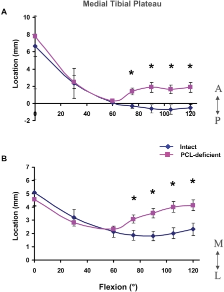
Fig. 3.
Location of peak cartilage deformation on the medial tibial plateau in the anteroposterior (A) and mediolateral (B) directions in the intact and posterior cruciate ligament (PCL)-deficient knees as a function of knee flexion angle. A = anterior to mediolateral axis, P = posterior to mediolateral axis, M = medial to anteroposterior axis, and L = lateral to anteroposterior axis. The values are given as the mean and standard deviation. *P < 0.05 as determined with one-way repeated-measures analysis of variance.

