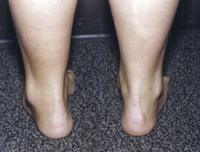
Fig. 1-A.
Figs. 1-A through 1-F Clinical photographs of a male patient with Charcot-Marie-Tooth disease who underwent reconstruction of the left foot at the age of eight years and eight months. In the preoperative photographs (Figs. 1-A, 1-C, and 1-E), the right foot appears to be relatively unaffected by deformity, while the left foot has pronounced hindfoot varus and cavus. The patient was lost to follow-up but returned to the clinic twelve years later, at which time he requested surgery for the right foot. Photographs made at that time (Figs. 1-B, 1-D, and 1-F) show slight recurrence of hindfoot varus and excellent maintenance of the cavus correction of the left (operatively treated) foot and substantial progression of the right (untreated) foot deformity.

