Abstract
Background: The geometry of the tibial plateau is complex and asymmetric. Previous research has characterized subject-to-subject differences in the tibial plateau geometry in the sagittal plane on the basis of a single parameter, the posterior slope. We hypothesized that (1) there are large subject-to-subject variations in terms of slopes, the depth of concavity of the medial plateau, and the extent of convexity of the lateral plateau; (2) medial tibial slope and lateral tibial slope are different within subjects; (3) there are sex-based differences in the slopes as well as concavities and convexities of the tibial plateau; and (4) age is not associated with any of the measured parameters.
Methods: The medial, lateral, and coronal slopes and the depth of the osseous portion of the tibial plateau were measured with use of sagittal and coronal magnetic resonance images that were made for thirty-three female and twenty-two male subjects, and differences between the sexes with respect to these four parameters were assessed. Within-subject differences between the medial and lateral tibial slopes also were assessed. Correlation tests were performed to examine the existence of a linear relationship between various slopes as well as between slopes and subject age.
Results: The range of subject-to-subject variations in the tibial slopes was substantive for males and females. However, the mean medial and lateral tibial slopes in female subjects were greater than those in male subjects (p < 0.05). In contrast, the mean coronal tibial slope in female subjects was less than that in male subjects (p < 0.05). The correlation between medial and lateral tibial slopes was poor. The within-subject difference between medial and lateral tibial slopes was significant (p < 0.05). No difference in medial tibial plateau depth was found between the sexes. The subchondral bone on the lateral part of the tibia, within the articulation region, was mostly flat. Age was not associated with the observed results.
Conclusions: The geometry of the osseous portion of the tibial plateau is more robustly explained by three slopes and the depth of the medial tibial condyle.
Clinical Relevance: The sex and subject-to-subject-based differences in the tibial plateau geometry found in the present study could be important to consider during the assessment of the risk of knee injury, the susceptibility to osteoarthritis, and the success of unicompartmental and total knee arthroplasty.
The articular surfaces of the tibiofemoral joint in combination with the primary ligaments play an important role in controlling the biomechanical behavior of the joint1,2. In particular, the contact mechanics between the femoral and tibial articular surfaces are complex, are three-dimensional, and consist of many unique features. Although we know a great deal about the three-dimensional geometry of the femoral condyles and their spherical profile for the portion of the femur that contacts the tibial plateau as the knee is flexed and extended3, very few quantitative data have been published regarding the asymmetric geometry of the tibial plateau. The geometry of the tibial plateau has a direct influence on the biomechanics of the tibiofemoral joint in terms of translation, the location of instantaneous center of rotation, the screw-home mechanism, and the strain biomechanics of the knee ligaments such as the anterior cruciate ligament1,2,4. An important characteristic of the tibial plateau is its posterior slope (with the anterior elevation being higher than the posterior elevation); when this characteristic is considered in association with a large compressive joint-reaction force such as that produced during weight-bearing activities, the force may have an anteriorly directed shear force component that acts to produce a corresponding anteriorly directed translation of the tibia1,5. Tibial slope is defined as the angle between the perpendicular to the middle part of the diaphysis of the tibia and the line representing the posterior inclination of the tibial plateau6. This slope has been measured by various researchers1,6,7, and its importance to anterior tibial translation has been well established in both animal8,9 and human models1,5. During weight-bearing, as the slope of the tibial plateau increases, the magnitude of the anteriorly directed shear force component that is associated with the compressive joint force on the tibia also increases1,6,9,10.
The geometry of the tibial plateau also has been studied as an important factor in total knee arthroplasty11, anterior cruciate ligament injury7,12, and cases in which osteotomy is performed to restore normal alignment of the knee13,14. Other investigators have suggested that increasing the tibial slope could benefit posterior cruciate ligament-deficient knees, whereas decreasing it could benefit anterior cruciate ligament-deficient knees5.
Notwithstanding the importance of the shape (that is, the various slopes) of the tibial plateau, very little information is available in the literature to help one to understand or classify the subject-to-subject differences in the geometry of the tibial plateau. The main identifying measurement is the posterior slope as measured on lateral radiographs, on which the medial and lateral tibial plateaus are superimposed. However, the measured slope is a two-dimensional approximation of a complex, asymmetric, and three-dimensional surface; it ignores the differences in the medial and lateral aspects of the tibia. One way to address the asymmetric geometry of the tibial plateau is to measure the medial and lateral aspects of the plateau with use of magnetic resonance imaging. Matsuda et al. used this approach to measure and compare the posterior slopes of normal and varus knees in human subjects15. Furthermore, there are no measurements in the literature that address the lateral-medial slope of the tibial plateau in the coronal plane, the depth of the concavity of the medial tibial plateau, or the extent of convexity of the lateral plateau.
Materials and Methods
On the basis of an unblinded pilot study of twenty male and twenty female subjects that was approved by the Texas Tech University Health Sciences Center, we developed the plans, jointly with the University of Vermont, for a blinded study to measure and compare tibial slope data between sexes. A power analysis revealed the need for a minimum of twenty-two subjects of each sex to establish 80% power to protect against the undue rejection of the null hypothesis. Accordingly, a subsequent blinded study was performed after its approval by the institutional review boards at both institutions. Subjects were included if they had no evidence of substantial knee osteoarthritis, ligament injuries, meniscus lesions, or cartilage injuries. All subjects were skeletally mature. None of the subjects had undergone previous knee surgery. To this end, T1-weighted sagittal and coronal magnetic resonance imaging scans of the tibiofemoral joint were acquired for fifty-five subjects at the University of Vermont. The sample comprised twenty-two male and thirty-three female subjects. The identifying information of the subjects (name, age, sex, and the conditions of the visit) was deleted, and the recorded magnetic resonance images were sent to the Texas Tech University Health Sciences Center for analysis. The person analyzing the images (J.H.) did not have any information regarding the sex or age of the subjects. All measurements were made with use of the annotation tools on the digital Picture Archiving and Communication System provided by the University of Vermont. The software reports the angular values in single-digit integer format. This means that if the true measure of the angle is, for instance, 2.4°, the software rounds the value to 2°. On the other hand, if the actual angle is 2.6°, the software returns a value of 3°. Thus, the angles are measured with a sensitivity of 1° (−0.5° to 0.5°) and, for a given measurement, the maximum deviation from the actual value does not exceed 0.5°. The length measurements were made with a sensitivity of 0.1 mm.
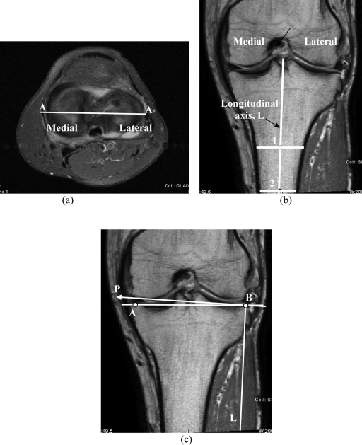
The procedure that was used to determine the tibial slope was based on the radiographic techniques developed by Genin et al.6. According to the procedure described by those authors, a lateral radiograph was analyzed by determining a line parallel to the middle part of the diaphysis of the tibia and then by constructing a line that was perpendicular to the middle part of the diaphysis and that passed through the tibial plateau. A second line representing the posterior slope of the tibia was then drawn. The angle between this line and the line perpendicular to the middle part of the diaphysis was considered to be the tibial slope. We applied the above procedure to magnetic resonance images as proposed by Matsuda et al.15. The first step was to identify a transverse plane passing through the tibiofemoral joint and showing the dorsal aspect of the tibial plateau (Fig. 1, a). In this transverse image, the coronal plane that passed closest to the centroid of the tibial plateau was identified. The location of this plane is represented by the horizontal line in Figure 1, a, and the corresponding coronal plane is shown in Figure 1, b. Next, the orientation of the longitudinal axis of the tibia, in the coronal plane, was determined. This was done by determining the midpoint (shown as small circles) of the medial-to-lateral width of the tibia at two points located approximately 4 to 5 cm apart and as distally in the image as possible (locations 1 and 2 in Figure 1, b). The extended line connecting the two midpoints represents the longitudinal (or diaphyseal) axis of the tibia in the coronal plane. The coronal slope of the tibial plateau (the coronal tibial slope) was then measured as the angle between a line joining the peak points on the medial and lateral aspects of the plateau (points A and B in Figure 1, c) and the line perpendicular to the longitudinal axis (line P in Figure 1, c). If point A fell below the perpendicular line P, then the slope was designated as positive (indicating that the peak lateral point was located proximal to the peak medial point).
Fig. 1.
Magnetic resonance images illustrating the method used to determine the coronal tibial slope. a: The transverse plane through the tibiofemoral joint shows the top view of the tibial plateau. The horizontal line AA represents the location of the coronal plane that was used to determine the diaphyseal axis. b: The orientation of the diaphyseal axis in the coronal plane. c: The coronal tibial slope (represented by the slope of line AB) is measured with respect to axis P, which is perpendicular to diaphyseal axis L.
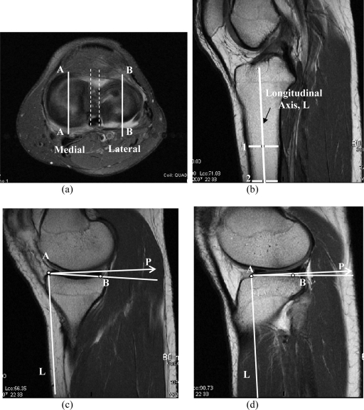
A similar approach was followed to determine the sagittal slopes of the medial and lateral tibial plateaus. In Figure 2, a, the solid vertical lines show the corresponding locations of the sagittal planes in the medial and lateral aspects of the tibial plateau. The dashed lines in the same figure show the approximate location of the sagittal plane that was used to determine the orientation of the diaphyseal or longitudinal axis. We used a plane that clearly showed the orientation of the tibia (Fig. 2, b). As before, the anterior and posterior cortices of the tibial shaft at two points located approximately 4 to 5 cm apart and distal in the image (points 1 and 2 in Figure 2, b) were determined. The midpoints of the lines representing the anterior-posterior thickness of the tibia were found, and the diaphyseal axis (L) was constructed. The diaphyseal axis was then reproduced in the medial plane as shown in Figure 2, c. The peak anterior and posterior points on the tibial plateau were identified (points A and B). The slope of the line extending through these two points represented the medial tibial slope, and it was measured with respect to the axis (P) perpendicular to the longitudinal axis (L). A similar approach was used to determine the lateral tibial slope (Fig. 2, d). Because the peak anterior points on the tibial plateau are proximal to the peak posterior points in both Figures 2, c and 2, d, the slopes of the medial tibial plateau and lateral tibial plateau would be positive according to the existing convention1,6.
Fig. 2.
Magnetic resonance images illustrating the method used to determine the medial and lateral tibial slopes. a: The vertical lines AA and BB represent the location of the sagittal plane used for determination of the medial and lateral tibial slopes. b: The sagittal plane (represented by dashed lines in Figure 2, a) was used to determine the orientation of the diaphyseal axis in the sagittal plane. c: The axis P perpendicular to L is reconstructed on the anterior peak of the medial tibial plateau, and the medial tibial slope (represented by the slope of line AB) is measured with respect to P. d: A similar approach is used to determine the lateral tibial slope.
The depth of the concavity of the medial aspect of the tibial plateau was measured by drawing a line connecting the superior and inferior crests of the tibial plateau in the same plane in which the medial tibial slope was measured (line AA in Figure 3, a). A line parallel to this line was then drawn tangential to the lowest point of the concavity, representing the lowest boundary of the subchondral bone (line BB in Figure 3, a). The perpendicular distance between the two lines (line CD in Figure 3, a) was then measured and was used to represent the depth of concavity of the medial tibial plateau. A similar approach was followed for the lateral side.
Fig. 3.
a: Magnetic resonance image illustrating the method used to determine the depth of the concavity of the medial aspect of the tibial plateau in the proximity of the center of the articulation region. b: Although the lateral aspect of the plateau (represented by ABCD) is convex overall, the articulation region (region BC) was quite flat in most subjects.
Once medial tibial slope, lateral tibial slope, and coronal tibial slope values were determined for all subjects, the tabulated data were mailed to the University of Vermont, where the subjects' age and sex codes were entered into the database. Student t tests were performed to compare the medial tibial slope, lateral tibial slope, and coronal tibial slope values between the sexes. A similar approach was used to compare the depths of tibial concavity between the sexes. Paired t tests were performed to test for differences between medial tibial slope and lateral tibial slope within individual subjects. Correlation analyses were performed to determine the strength of the linear relationships between medial tibial slope and lateral tibial slope, coronal tibial slope and medial tibial slope, and coronal tibial slope and lateral tibial slope. Finally, in a post hoc analysis, correlation tests were performed to assess the existence of a linear relationship between age and various slope measurements. The level of significance was set at 0.05 for all t tests and the correlation analysis.
Results
The means and standard deviations for medial tibial slope, lateral tibial slope, and coronal tibial slope are presented in Table I. For the male subjects, the medial tibial slope ranged from −3° to +10° and the lateral tibial slope ranged from 0° to +9°. For the female subjects, the medial tibial slope ranged from 0° to +10° and the lateral tibial slope ranged from +1° to +14°. The coronal tibial slope ranged from 0° to +6° for the male subjects and from −1° to +6° for the female subjects. The mean medial tibial slope for the female subjects was significantly greater than that for the male subjects (5.9° compared with 3.7°; p = 0.01). Similarly, the mean lateral tibial slope for the female subjects was significantly greater than that for the male subjects (7.0° compared with 5.4°; p = 0.02). There was a significant difference (pairwise comparison) between the within-subject medial tibial slope and lateral tibial slope values when the male and female subjects were considered as separate groups as well as when they were considered as a combined group (p < 0.05 for all comparisons). Finally, the mean coronal tibial slope for female subjects was significantly less than that for male subjects (2.5° compared with 3.5°; p = 0.03).
TABLE I.
Medial, Lateral, and Coronal Slopes of the Tibial Plateau for Male and Female Subjects and Corresponding P Values Establishing Differences Between Sexes
| Sagittal Tibial Slope
|
|||
|---|---|---|---|
| Medial Tibial Slope | Lateral Tibial Slope | Coronal Tibial Slope | |
| Female (n = 33) | |||
| Mean | 5.9° | 7.0° | 2.5° |
| Standard deviation | 3.0° | 3.1° | 1.9° |
| Male (n = 22) | |||
| Mean | 3.7° | 5.4° | 3.5° |
| Standard deviation | 3.1° | 2.8° | 1.9° |
| P value | 0.01 | 0.02 | 0.03 |
The reproducibility of the measurements was analyzed post hoc by randomly selecting fifteen subjects and measuring the tibial slopes in three independent trials (one original measurement as well as two additional independent measurements). The first trial (original measurement) and the second trial were separated by three months, whereas the second trial and the third trial were separated by four days. We then performed an intraclass correlation analysis to assess the extent of the reproducibility16. The analysis revealed a significant (p < 0.05) correlation coefficient of 0.88, demonstrating that the intraobserver reproducibility was acceptable. The estimate of within-subject variance was 0.67 degrees squared, and the estimate of between-subject variance was 7.4 degrees squared.
The depth of the subchondral bone concavity of the medial compartment of the tibia ranged from 1.4 to 4.2 mm (mean, 2.7 ± 0.76 mm) for female subjects and from 1.2 to 5.2 mm (mean, 3.1 ± 0.99 mm) for male subjects. There was no difference in the depth of concavity of the medial compartment between the male and female subjects (p = 0.1); however, the statistical power associated with this comparison was low (34%). The extent of the convexity of the lateral compartment of the tibia was not large enough for a meaningful measurement. On the lateral side, although the osseous boundary of the tibial plateau is clearly convex (as represented by boundary ABCD in Figure 3, b), the subchondral bone directly distal to the femur (the region over which the femoral condyle articulates on the lateral tibia) is predominantly flat (region BC in Figure 3, b).
The female subjects ranged in age from thirteen to sixty-four years (mean and standard deviation, 33.2 ± 15.3 years), whereas the male subjects ranged from fourteen to fifty-six years (mean and standard deviation, 34.6 ± 11.0 years). In a post hoc analysis, all correlation coefficients, including medial tibial slope-age, lateral tibial slope-age, and coronal tibial slope-age for both of the individual sexes as well as the pooled populations, were <0.25.
The correlation coefficients between medial tibial slope and lateral tibial slope, coronal tibial slope and medial tibial slope, and coronal tibial slope and lateral tibial slope for the pooled population were not strong, as evidenced by their values of 0.5, −0.26, and −0.27, respectively; similar results were found for comparisons in the individual sex populations.
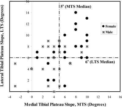
In an effort to provide added insight into the differences in the tibial slopes between the sexes, the medial tibial slope (abscissa) and lateral tibial slope (ordinate) values from all subjects were plotted on the same graph (Fig. 4). The graph was then divided into four regions by two lines representing the median values, for the pooled population, for medial tibial slope (indicated by the vertical dotted line at 5°) and lateral tibial slope (indicated by the horizontal dotted line at 6°). In Figure 4, it is interesting to note that the data points for a substantial proportion of the female subjects (36%) were located in the top right quadrant, with high medial tibial slope and lateral tibial slope values, whereas the data points for a substantial proportion of male subjects (31%) appeared in the lower left quadrant, with lower medial tibial slope and lateral tibial slope values. It is also apparent that the data points for a small proportion of the subjects were located in the top left quadrant, where medial tibial slope is considerably smaller than lateral tibial slope, and that the data points for an even smaller proportion of the subjects, both male and female, were located in the lower right quadrant, where medial tibial slope is considerably larger than lateral tibial slope. More importantly, 33% of the female subjects had a medial tibial slope of ≥8°, compared with only 10% of the male subjects. Similarly, 27% of the female subjects had a lateral tibial slope of ≥9°, compared with 10% of the male subjects.
Fig. 4.
Graph showing the medial tibial slope (MTS) and lateral tibial slope (LTS) values for male and female subjects. The data points for female subjects are concentrated in the top right quadrant, indicating the highest medial tibial slope and lateral tibial slope values.
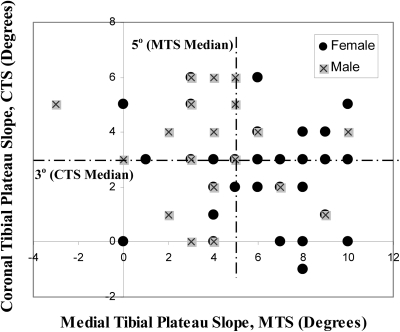
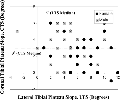
Similar plots were constructed with use of the medial tibial slope and coronal tibial slope data (Fig. 5) and the lateral tibial slope and coronal tibial slope data (Fig. 6). These graphs were also divided into four quadrants with use of the medians of the pooled populations. Figure 5 shows that, with exclusion of the median values, the data points for the greatest number of the female subjects were located in the bottom right quadrant (27%) whereas the data points for the male subjects had a more homogeneous distribution over the entire plane (with 9% being located in the bottom right quadrant). Additionally, with exclusion of the median values, the data points for 54% of the female subjects fell below the coronal tibial slope median of 3°, compared with those for only 27% of the male subjects. Similarly, in Figure 6, the data points for the majority of female subjects were distributed on the right-hand side of the plane, whereas the data points for male subjects were more homogeneously distributed.
Fig. 5.
Graph showing the medial tibial slope (MTS) and coronal tibial slope (CTS) values for male and female subjects. The data points for female subjects are concentrated in the bottom right quadrant of the plane, indicating lower coronal tibial slope and higher medial tibial slope combinations.
Fig. 6.
Graph showing the lateral tibial slope (LTS) and coronal tibial slope (CTS) values for male and female subjects. The data points for female subjects are concentrated in the bottom right quadrant of the plane, indicating lower coronal tibial slope and higher lateral tibial slope combinations.
Discussion
Much of what is known about the geometry of the asymmetric, three-dimensional, osseous portion of the tibial plateau is based on two-dimensional measurements obtained from lateral radiographs. With that approach, it is difficult to differentiate between the medial and lateral aspects of the plateau because they are superimposed. The true tibial slope should be based on measurements made at the center of the articular regions of the medial and lateral compartments of the tibial plateau. The approach used in the present study was based on the accepted method of measuring the tibial slope on radiographic images1,6 but involved the use of magnetic resonance images instead15, which allowed us to characterize the slope of the tibial plateau at the center of the articular surfaces of the medial and lateral compartments. Furthermore, the measurement of the depth of the concavity of the medial compartment adds another dimension to our measurements that helps to characterize the complex three-dimensional geometry of the tibial plateau. If one compares knees with similar articular cartilage thickness profiles and meniscal geometries, a deeper medial plateau will constrain the femoral condyle to a greater extent and may result in increased resistance to displacement of the tibia relative to the femur. Conversely, the combination of a high medial tibial slope and low depth of concavity may be associated with a decreased resistance to displacement of the tibia relative to the femur, placing the knee at increased risk for ligament injury (for example, anterior cruciate ligament tears). Our results show that although the lateral aspect of the plateau is convex, the subchondral bone beneath the articular region is predominantly flat (Fig. 3, b). Therefore, the posteriorly directed slope of the lateral aspect of the tibial plateau may be sufficient to explain its contribution to the biomechanics of the tibiofemoral joint.
We found that there were significant differences in the medial and lateral slopes of the tibial plateau between the male and female subjects and that age was not associated with these differences. The differences, while significant, were not large, particularly for the coronal tibial slope comparisons. We evaluated our ability to detect such small differences with the accuracy of the measurement technique that was used. The accuracy of each measurement was considered to be a random variable that was uniformly distributed in the interval (a, b). In our case, this interval was 1° (−0.5° to 0.5°). If we denote a measurement plus its accuracy error by Y, then  , where
  is the variance of the accuracy error and
  is the variance of the actual slopes. For a uniform distribution where every measurement is subjected to the same accuracy error17, the variance is
  . Thus, in our case,
  degrees squared. We therefore note that the effect of measurement accuracy is very small in comparison with the estimates of
  (note values of standard deviation in Table I) as in each case
  . Thus, the established differences are significant.
When the data from all subjects were considered, we also found that for both the medial and lateral compartments there were a large range of slopes, extending from −3° to +10° for medial tibial slope, from 0° to +14° for lateral tibial slope, and from −1° to +6 ° for coronal tibial slope. These ranges are similar to those observed by Matsuda et al.15, who reported a range of 5° to 15.5° for medial tibial slope and a range of 0° to 14.5° for lateral tibial slope in their study of subjects with normal knees. Additionally, the two reported mean values for lateral tibial slope in our combined sample (6.38° ± 3.04°) and the sample in the study by Matsuda et al.15 (7.2° ± 3.8°) are similar. In contrast, the mean values of the medial tibial slope measurements differ (5.02° ± 3.2° in the present study, compared with 10.7° ± 2.4° in the study by Matsuda et al.15). The most dramatic differences were observed in the maximum values of the medial tibial slope (with the value in the study by Matsuda et al.15 being 5.5° greater than that in our study) and the minimum values of the medial tibial slope (with the value in the study by Matsuda et al.15 being 8° greater than that in our study). These findings may be attributed to differences in the choice of the reference axes used for measurement of the tibial slopes, differences in the distribution of outliers in our study as compared with that in the study by Matsuda et al.15, and the unknown distribution of sexes in the study by Matsuda et al.15. A review of our data reveals that almost any combination of slopes in the above ranges could exist and that a strong correlation did not exist between any of the measured variables (Figs. 4, 5, and 6). Thus, it is possible that the study by Matsuda et al.15 included more females in the healthy group, resulting in higher medial tibial slope values.
The two previous studies in which sex-based comparisons of tibial slope were made involved the use of a conventional radiographic approach and did not demonstrate a significant difference7,13. The medial and lateral slopes of the tibia were not measured in either study. In our study, female subjects had a steeper posteriorly directed tibial slope medially (by an average of 59%) and laterally (by 30%) in comparison with male subjects. In contrast, the coronal tibial slope, which is oriented downward in a lateral-to-medial direction, was less steep in female subjects (by 29%). The similarity of standard deviations in the male and female groups indicates that the degree of scatter about the means is similar (Table I). Thus, characterizing the entire tibial plateau on the basis of a single slope is inadequate. It is important to note that the averages and standard deviations reported here for both the medial tibial slope and the lateral tibial slope for the pooled population (5.02° ± 3.2° and 6.38° ± 3.04°, respectively) are considerably less than those reported previously1,7 (10° ± 3° and 8.8° ± 1.8°, respectively). We believe that this difference is explained by our use of a magnetic resonance imaging-based approach instead of a radiographic approach and the choice of the reference axes used to measure the tibial slopes.
The slope of the tibial plateau has a direct relationship with anterior tibial translation of the knee as it transitions from non-weight-bearing to weight-bearing conditions1,9,18,19. This relationship is supported by the findings of Torzilli et al.18, who used a cadaver model to demonstrate that the application of a compressive load to the knee produces an anteriorly directed shear force that results in an anterior neutral position shift of the tibia relative to the femur. The magnitude of this displacement was greater in anterior cruciate ligament-sectioned knees than in intact knees. Our research group has reported similar findings in humans19. The amount of anterior translation produced during the transition from non-weight-bearing to weight-bearing is not only produced by the orientation of the patellar tendon relative to the tibial plateau, it is also influenced by the magnitudes of the compressive joint load and the posteriorly directed slope of the tibial plateau. During transmission of a compressive force across the tibiofemoral joint, an increase in the slope of the tibial plateau is associated with an increase in the magnitude of the anteriorly directed force component of the compressive force that acts on the articular surface of the tibia, a force that acts to produce a corresponding anteriorly directed translation of the tibia. This relationship has been explained with use of mathematical models that have shown that the resultant anterior cruciate ligament force increases as the slope of the tibial plateau increases20,21. It has been reported that, during the early stance phase of gait, subjects with anterior cruciate ligament-deficient knees and a 4° slope of the tibia experienced a 9.1-mm anterior translation of the tibia relative to the femur, whereas those with a 12° slope experienced 15.2 mm of anterior translation22. It can be argued that because females have a steeper tibial slope in both the medial and lateral compartments, the application of large compressive joint loads, such as those produced during the transition from non-weight-bearing to weight-bearing postures, exposes them to greater magnitudes of anteriorly directed force on the proximal part of the tibia, and this may predispose them to an increased risk of anterior cruciate ligament injury in comparison with males. If one considers the mean values of tibial slope, it could be argued that the mean differences between females and males of 2.2°, 1.6°, and 1.0° in medial tibial slope, lateral tibial slope, and coronal tibial slope may not have a dramatic effect on the biomechanics of the knee5; however, the important point to consider is the differences between the extreme values. For example, 33% of the female subjects had a medial tibial slope of ≥8° and 27% had a lateral tibial slope of ≥9°. Conversely, only 10% of the male subjects had a medial tibial slope of ≥8° and 10% had a lateral tibial slope of ≥9°. The concentration of the data points for female subjects in the top right quadrant of Figure 4 compared with the clustering of the data points for male subjects in the lower left quadrant demonstrates this difference. Thus, one can argue that the anteriorly directed forces on the tibia that result from the compressive joint load applied to the tibiofemoral joint, which must be resisted by the anterior cruciate ligament, vary considerably when one considers a medial tibial slope of 10° in a female subject as opposed to a medial tibial slope of 0° in a male subject (or even another female subject). From this perspective, Figures 4, 5, and 6 are important because they present information regarding the distribution of the medial tibial slope, lateral tibial slope, and coronal tibial slope measurements for male and female subjects. Furthermore, these figures show the subject-to-subject differences in the range and distribution of these tibial slopes. In reality, these figures show that differences between the sexes, although important, are less important than differences between the subjects.
The lateral-to-medial slope of the tibial plateau, or the coronal tibial slope, was positive or zero for 98% of the subjects. There was only one subject with a coronal tibial slope of −1°, and the remaining subjects had a coronal tibial slope that ranged between 0° and 6° (Figs. 5 and 6). This finding indicates that the lateral edge is superior to the medial edge of the tibial plateau in a majority of subjects. However, this slope is significantly larger in male subjects as evidenced by the distribution presented in Figures 5 and 6. In general, males have a higher coronal tibial slope but a lower medial tibial slope and lateral tibial slope than females. Although the effect that coronal tibial slope has on the biomechanics of the tibiofemoral joint is not entirely understood at this time, one could envision an influence on the mechanics of anterior cruciate ligament injury (both contact and noncontact) and on the ligament balancing associated with unicompartmental knee reconstruction in which only one aspect of the joint is reconstructed and the maintenance of normal knee biomechanics may be a desired clinical goal.
The findings of the present study suggest that future studies should consider the three tibial slopes (medial tibial slope, lateral tibial slope, and coronal tibial slope) as well as the depth of concavity of the medial plateau as potential predictors of noncontact anterior cruciate ligament injury. The data presented in Figures 4, 5, and 6 could serve as a basis to distinguish at-risk tibial geometries, and the same convention could be used to determine at-risk geometries for the onset and progression of osteoarthritis.
Furthermore, in both total and unicondylar knee arthroplasty, understanding the subject-to-subject differences in terms of medial tibial slope, lateral tibial slope, and coronal tibial slope could lead to a more accurate subject-specific implant that replicates the biomechanics of the normal knee11,23. Improvements may be possible if the surgeon attempts to place the prosthetic tibial component at a specific slope that reproduces the original compartmental slope of the subject.
Finally, the knee laxity measurements that are often measured at 15° to 25° of knee flexion with use of an arthrometer may be influenced by the tibial slope as well as the medial tibial depth. Differences in medial tibial slope, lateral tibial slope, and the depth of concavity of the medial aspect of the tibial plateau could influence the resistance of the tibia to anterior translation, and this could influence the anterior knee laxity capacity that is measured with use of arthrometers. Future studies may be needed to assess the correlation between tibial slope and anterior knee laxity measurements.
One of the limitations of the present study is that we did not have access to the height and weight of the subjects, and consequently we did not explore correlations that may exist with the measured slopes. These covariates should be considered in future research regarding the role of tibial slope in anterior cruciate ligament injury and osteoarthritis. As a second limitation, it is important to note that the magnetic resonance imaging voxel resolution, the access to a sufficient length of the tibia in the magnetic resonance image, and the ability to identify landmarks precisely all could have an impact on the slope measurements. However, although these factors may influence the results, they will not influence the large subject-to-subject differences or the large range of slope values described in the present report.
In conclusion, the present study introduces a new approach to describe the slopes of the medial and lateral aspects of the osseous portion of the tibial plateau in the sagittal and coronal planes. It is more comprehensive and relays more information regarding the biomechanics of the tibiofemoral joint in comparison with previous reports. These geometric differences may be important to consider when assessing the risk of knee injury, the susceptibility to osteoarthritis, and the design and success of unicondylar and total knee arthroplasty. 
Acknowledgments
Note: The corresponding author (J.H.) acknowledges the support of Texas Tech College of Engineering and Texas Tech Health Sciences Center. The authors thank Dr. Evelyne Fliszar, University of Vermont, for her assistance in the collection and evaluation of the magnetic resonance imaging images. The support of the National Institutes of Health (NIAMS/NIH), Grant Number R01AR050421 (Beynnon BD) is gratefully achnowledged.
Disclosure: In support of their research for or preparation of this work, one or more of the authors received, in any one year, outside funding or grants in excess of $10,000 from the National Institutes of Health (NIAMS/NIH), Grant Number R01AR050421. Neither they nor a member of their immediate families received payments or other benefits or a commitment or agreement to provide such benefits from a commercial entity. No commercial entity paid or directed, or agreed to pay or direct, any benefits to any research fund, foundation, division, center, clinical practice, or other charitable or nonprofit organization with which the authors, or a member of their immediate families, are affiliated or associated.
Investigation performed at Texas Tech University, Lubbock, Texas, and the University of Vermont, Burlington, Vermont
References
- 1.Dejour H, Bonnin M. Tibial translation after anterior cruciate ligament rupture. Two radiological tests compared. J Bone Joint Surg Br. 1994;76:745-9. [PubMed] [Google Scholar]
- 2.Beynnon B, Yu J, Huston D, Fleming B, Johnson R, Haugh L, Pope MH. A sagittal plane model of the knee and cruciate ligaments with application of a sensitivity analysis. J Biomech Eng. 1996;118:227-39. [DOI] [PubMed] [Google Scholar]
- 3.Coughlin KM, Incavo SJ, Churchill DL, Beynnon BD. Tibial axis and patellar position relative to the femoral epicondylar axis during squatting. J Arthroplasty. 2003;18:1048-55. [DOI] [PubMed] [Google Scholar]
- 4.Nordin M, Frankel VH. Basic biomechanics of the musculoskeletal system. 3rd ed. Philadelphia: Lippincott Williams and Wilkins; 2001.
- 5.Giffin JR, Vogrin TM, Zantop T, Woo SL, Harner CD. Effects of increasing tibial slope on the biomechanics of the knee. Am J Sports Med. 2004;32:376-82. [DOI] [PubMed] [Google Scholar]
- 6.Genin P, Weill G, Julliard R. [The tibial slope. Proposal for a measurement method]. J Radiol. 1993;74:27-33. French. [PubMed] [Google Scholar]
- 7.Meister K, Talley MC, Horodyski MB, Indelicato PA, Hartzel JS, Batts J. Caudal slope of the tibia and its relationship to noncontact injuries to the ACL. Am J Knee Surg. 1998;11:217-9. [PubMed] [Google Scholar]
- 8.Slocum B, Devine T. Cranial tibial thrust: a primary force in the canine stifle. J Am Vet Med Assoc. 1983;183:456-9. [PubMed] [Google Scholar]
- 9.Slocum B, Devine T. Cranial tibial wedge osteotomy: a technique for eliminating cranial tibial thrust in cranial cruciate ligament repair. J Am Vet Med Assoc. 1984;184:564-9. [PubMed] [Google Scholar]
- 10.Dejour H, Walch G, Chambat P, Ranger P. Active subluxation in extension. Am J Knee Surg. 1988;1:204-11. [Google Scholar]
- 11.Massin P, Gournay A. Optimization of the posterior condylar offset, tibial slope, and condylar roll-back in total knee arthroplasty. J Arthroplasty. 2006;21:889-96. [DOI] [PubMed] [Google Scholar]
- 12.Brandon ML, Haynes PT, Bonamo JR, Flynn MI, Barrett GR, Sherman MF. The association between posterior-inferior tibial slope and anterior cruciate ligament insufficiency. Arthroscopy. 2006;22:894-9. [DOI] [PubMed] [Google Scholar]
- 13.DeJour H, Neyret P, Bonnin M. Instability and osteoarthritis. In: Fu FH, Harner CD, Vince GV, editors. Knee surgery. Baltimore: Lippincott Williams and Wilkins; 1994. p 859-76.
- 14.Rodner CM, Adams DJ, Diaz-Doran V, Tate JP, Santangelo SA, Mazzocca AD, Arciero RA. Medial opening wedge tibial osteotomy and the sagittal plane: the effect of increasing tibial slope on tibiofemoral contact pressure. Am J Sports Med. 2006;34:1431-41. [DOI] [PubMed] [Google Scholar]
- 15.Matsuda S, Miura H, Nagamine R, Urabe K, Ikenoue T, Okazaki K, Iwamoto Y. Posterior tibial slope in the normal and varus knee. Am J Knee Surg. 1999;12:165-8. [PubMed] [Google Scholar]
- 16.Shrout PE, Fleiss JL. Intraclass correlations: uses in assessing rater reliability. Psych Bull. 1979;86:420-7. [DOI] [PubMed] [Google Scholar]
- 17.Hogg RV, McKean JW, Craig AT. Introduction to mathematical statistics. 6th ed. Upper Saddle River, NJ: Pearson Education; 2005.
- 18.Torzilli PA, Deng X, Warren RF. The effect of joint-compressive load and quadriceps muscle force on knee motion in the intact and anterior cruciate ligament-sectioned knee. Am J Sports Med. 1994;22:105-12. [DOI] [PubMed] [Google Scholar]
- 19.Beynnon BD, Fleming BC, Labovitch R, Parsons B. Chronic anterior cruciate ligament deficiency is associated with increased anterior translation of the tibia during the transition from non-weightbearing to weightbearing. J Orthop Res. 2002;20:332-7. [DOI] [PubMed] [Google Scholar]
- 20.Chan SC, Seedhom BB. The effect of the geometry of the tibia on prediction of the cruciate ligament forces: a theoretical analysis. Proc Inst Mech Eng [H]. 1995;209:17-30. [DOI] [PubMed] [Google Scholar]
- 21.Imran A, O'Connor JJ. Theoretical estimates of cruciate ligament forces: effects of tibial surface geometry and ligament orientations. Proc Inst Mech Eng [H]. 1997;211:425-39. [DOI] [PubMed] [Google Scholar]
- 22.Liu W, Maitland ME. Influence of anthropometric and mechanical variations on functional instability in the ACL-deficient knee. Ann Biomed Eng. 2003;31:1153-61. [DOI] [PubMed] [Google Scholar]
- 23.Aleto TJ, Berend ME, Ritter MA, Faris PM, Meneghini RM. Early failure of unicompartmental knee arthroplasty leading to revision. J Arthroplasty. 2008;23:159-63. [DOI] [PubMed] [Google Scholar]


