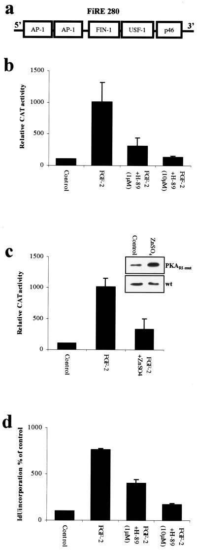
Figure 1.
PKA is required for FGF-2-induced activation of FiRE and induction of cell proliferation. (a) Schematic model of the FGF-inducible response element (FiRE), located at −10 kb of the translation initiation site of murine syndecan-1 gene. When activated by FGF-2, FiRE binds AP-1, a 50-kDa FIN-1, and USF-1 transcription factors. (b) PKA-specific inhibitor H-89 inhibits the FGF-2-induced activation of FiRE in a concentration-dependent manner. NIH 3T3 cells were stably transfected with FiRE-CAT plasmid, serum-starved for 48 hr, and treated for 30 min with H-89 (1 μM or 10 μM) before overnight exposure to FGF-2 (10 ng/ml), followed by determination of CAT activity. Means and standard deviations of three independent experiments of three parallel wells are shown in each. (c) The expression of dominant-negative PKARΙ inhibits the FGF-2-induced activation of FiRE. NIH 3T3 cells bearing the p271FiRECAT construct were transfected by a construct encoding a dominant-negative form of PKARΙ (PKARΙ-mut). The production of PKARΙ-mut was induced by application of ZnSO4 (50 μM final concentration) into the culture medium 3 hr before a 12-hr FGF-2 stimulation. Means and standard deviations of three independent experiments are shown. Control represents CAT activity from non-growth factor-treated cells. The ZnSO4-induced production of the dominant-negative PKARΙ was verified with anti-PKARΙ antibody. PKARΙ-mut stably transfected cells were treated with 50 μM ZnSO4 for 12 hr, and protein lysates were collected, blotted, and detected with anti-PKARΙ. As a control, nontransfected NIH 3T3 cells (wt) were similarly treated with ZnSO4. The immunoblots show significant increase in total PKARΙ immunoreactivity in ZnSO4-treated stably transfected cells, whereas in nontransfected cells, ZnSO4 treatment had no effect on the amount of PKARΙ. (d) PKA-specific inhibitor H-89 inhibits the FGF-2-induced DNA synthesis of NIH 3T3 cells in a concentration-dependent manner. Serum-starved NIH 3T3 cells were pretreated with H-89 (1 μM or 10 μM) for 30 min before an 18-hr FGF-2 treatment, and the incorporated radioactivity was measured with a γ counter.

