
Figure 2.
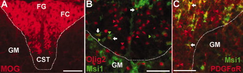
Msi1 expression in the mouse spinal cord during active myelination. Transverse sections through the dorsal funiculus white matter of lumbar spinal cords from P7 mice. A dashed line demarcates the white matter of the dorsal funiculus from the adjacent gray matter (GM) of the dorsal horns. A: MOG immunolabeling illustrates ongoing myelination within the tracts of the dorsal funiculus. The fasciculus cuneatus (FC) has relatively extensive MOG immunolabeling while the area of the fasciculus gracilis (FG) is incompletely immunolabeled and most of the ventral area corresponding with the corticospinal tract (CST) has not yet acquired myelin immunolabeling. B: Immunohistochemistry for Msi1 expression (green) with Olig2 (red), as a marker of oligodendrocyte lineage cells, reveals double labeled Msi1+ Olig2+ cells (B, white arrows) among many Msi1+ Olig2‐ cells (green arrowheads) and Msi1‐ Olig2+ cells (red arrowheads). C: Immunohistochemistry for Msi1 expression (green) and PDGFαR (red), as a marker of OP cells, demonstrates a subset of Msi1+ PDGFαR+ cells (C, white arrows) among many Msi1+ PDGFαR‐ cells (green arrowheads) and Msi1‐ PDGFαR+ cells (red arrowheads). Scale bars = 50 μm.