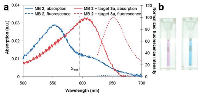
Figure 2.

(a) Absorption (solid) and fluorescence emission (dashed, λexc) = 605 nm) spectra of SQuID MB 2 (1.49 μM) with (red) and without (blue) oligonucleotide 3a (19× excess) present. In the absence of target, the shoulder absorption at 605 nm is due to a fraction of molecules not in the H-dimer configuration. (b) Photograph of SQuID MB 2 solutions before (left) and after (right) target 3a addition showing obvious color change.
