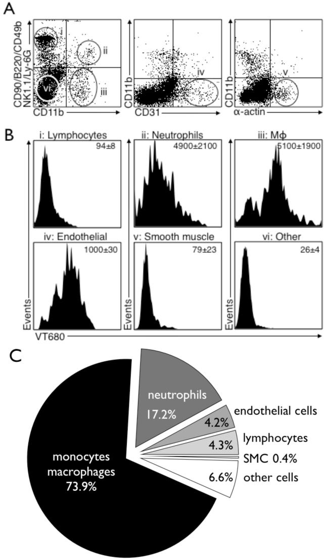
Figure 6. Flow cytometry analysis of digested atherosclerotic lesions shows predominant uptake of 64Cu-TNP into monocyte/macrophages.
Flow cytometry was performed on digested aortas of apoE-/- injected with the nanoparticle.
6A: Placement of gates within all living cells: i) lymphocyte gate, ii) neutrophil gate, iii) Mø gate, iv) endothelial cell gate, v) smooth muscle cell gate, and vi) other cell types.
6B: Histograms with respective intracellular signal in the VT680 channel for each cell type as identified in 6A.
6C: Relative signal contribution per cell type to overall signal. Mø are the dominating cell type ingesting 64Cu-TNP (73.9% of overall signal), followed by neutrophils (17.2%), endothelial cells (4.2%), lymphocytes (4.3%) and smooth muscle cells (0.4%) and other cell types not stained by the used antibody cocktail (6.6%).

