
FIG. 2.
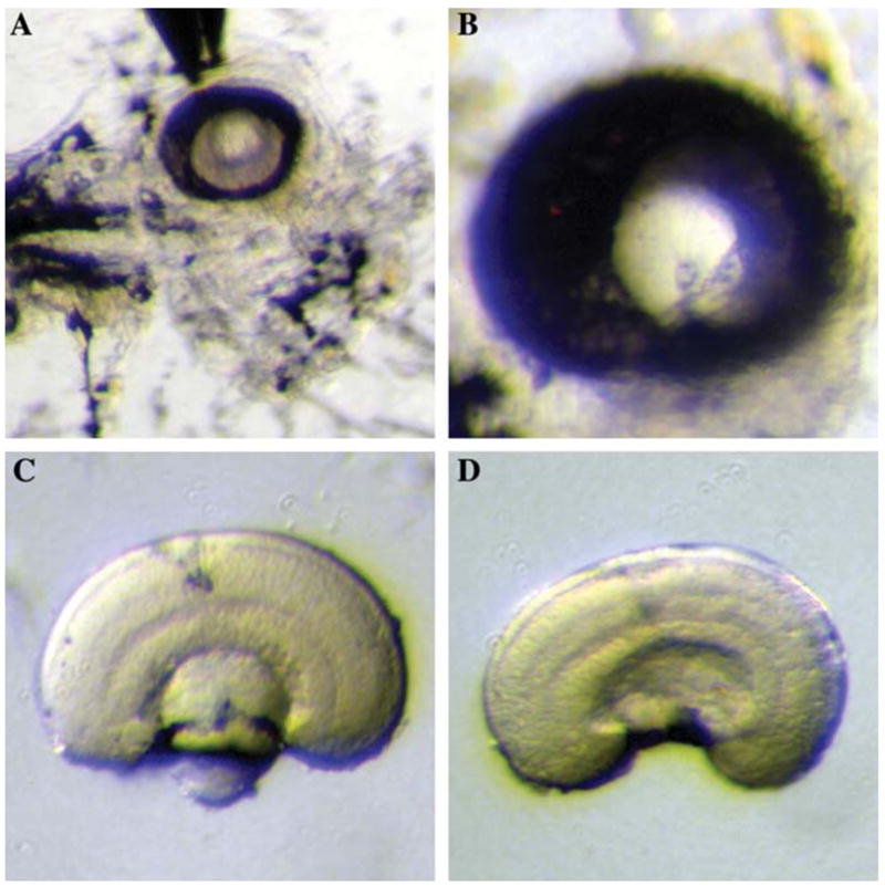
Dissection of zebrafish embryonic retina. (A) The removal of RPE. The anterior side of the embryo is facing right and the dorsal side is facing upward. The RPE is partially opened and cleaned up from the medial side. (B) A higher magnification of the eye with the same orientation as in (A), with RPE partially cleaned-up. (C) Dissected retina before the removal of the lens, observed from the side. (D) Dissected retina after the removal of the lens, observed from the side. Note that the cell and plexiform layers, as well as the optic nerve, are apparent after the RPE is peeled off.