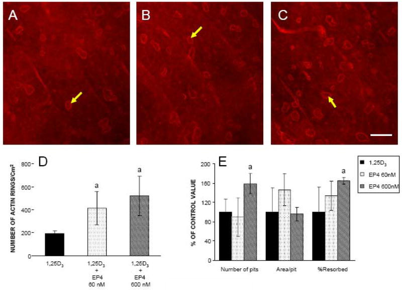
Figure 7.
Effects of EP4 and 1,25D3 on the formation of actin rings and resorptive pits on bone slices. Marrow cells that had been cultured for 5 days with 1,25D3 were scraped and plated on bone slices. Pits were determined by examining bone slices using differential interference contrast optics. A. Cultures treated with 1,25D3 alone, B. Cultures treated with 60 nM EP4 + 1,25D3, C. Cultures treated with 600 nM EP4 +1,25D3 with arrows showing the actin rings. Scale bar = 25 μm, D. Quantitation of the actin rings showing that their formation in response to EP4 is dose dependent, E. Quantitation of the pitting is shown in the bar graph. Values are expressed as the mean ± SD of 5 measurements. aSignificantly different from culture treated with 1,25D3 (P<0.05).

