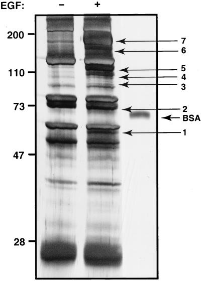
Figure 1.
EGF-induced tyrosine phosphorylation in HeLa cells. Serum-deprived HeLa S3 cells (5 × 10⁹) were either left untreated or treated with 1 μg/ml EGF for 5 min. Cleared cell lysates were immunoprecipitated with a mixture of monoclonal anti-phosphotyrosine antibodies, washed, and resolved by SDS/PAGE. The gel was then silver-stained. Molecular mass markers in kDa are indicated as well as 150 fmol of BSA that was loaded onto the same gel. The arrows with numbers indicate the positions of the bands that were excised for enzymatic digestion by trypsin and subsequent mass spectrometric analysis.

