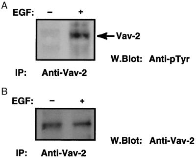
Figure 3.
Vav-2 is tyrosine phosphorylated by EGF treatment. HeLa cells were either left untreated or treated with EGF for 5 min, and lysates were immunoprecipitated (IP) with anti-Vav-2 antibody as indicated. Washed immunoprecipitates were resolved by SDS/PAGE, transferred onto nitrocellulose, and Western blotted with an anti-phosphotyrosine antibody (A). The position of tyrosine-phosphorylated Vav-2 is indicated by an arrow. B shows equal loading of Vav-2 from a parallel experiment.

