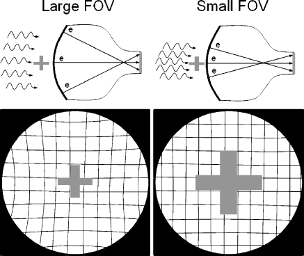
Fig. 2.
Electronic focusing allows the II to change the FOV and magnify the anatomy. The upper and lower left drawings illustrate the II/TV in the large FOV mode, providing good coverage and high gain as a result of electron minification. Note the presence of geometric ‘pin-cushion–distortion in the periphery of the image. This is caused by mapping the spherical input phosphor electron image onto the planar output phosphor. In magnification mode (smaller FOV), shown in the upper and lower right drawings, objects are magnified and spatial resolution is improved; however, this is at the expense of lower gain (less minification) and higher patient dose. Geometric pin-cushion distortion is reduced as there is less ‘warping–of the image onto the output phosphor from the central area of the input phosphor

