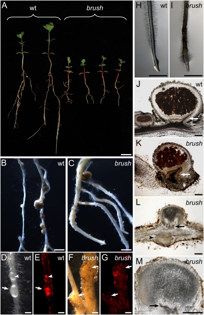
Figure 1.
brush shoot, root, and nodulation mutant phenotypes. A, brush mutant plants display shorter roots and shoots in comparison to wild-type (wt) plants. Stereomicroscopic images of wild-type (B, D, and E) and brush (C, F, and G) roots with young developing nodules (arrows) are shown in bright-field (B, C, D, and F) and red fluorescence emission (E and G) microscopy. Inside wild-type nodules, a strong fluorescent signal (E, arrow) and invading ITs (D and E, arrowheads) could be detected, whereas mutant nodule structures (F and G) lack bacterially expressed red fluorescence inside the bump-like structures that were initiated on the root (arrows). Stereomicroscopic images of wild-type (H) and brush (I) roots of plants grown in the presence of 0.01 μm AVG for 1 week on agar plates at 26°C. Bright-field microscopic imaging of 40-μm sections of brush versus wild-type mature and immature nodules. J, Section of wild-type immature and mature nodules. K, Section of infected mature brush nodule. L, Longitudinal section of the typical brush aberrant root structure with a nodule and a nodule primordium without bacterial infection (arrowhead), which did not develop any nodule vascular bundle at the base. M, Mature, uninfected brush nodule. Arrows in J to M indicate the nodule vascular bundle. B to G and J to M show nodules from plants that were grown for 4 weeks in the presence of M. loti MAFF303099 (DsRed) in glass jars at 26°C. Scale bars: 1 cm (A); 2 mm (B, C, H, and I); 200 μm (D–G, J–M).

