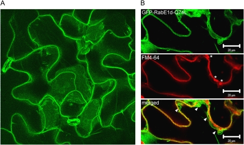
Figure 4.
Intracellular localization of the mutant GFP- RabE1d-Q74L protein. A, Confocal microscopy image of a representative Arabidopsis leaf expressing GFP-RabE1d-Q74L. Projection along the z axis of several focal planes crossing the epidermal cell layer. Several independent transgenic lines were analyzed with similar results. B, GFP-RabE1d-Q74L is mostly localized in the tonoplast. Leaves were stained with FM4-64, to visualize the plasma membrane, and immediately observed. The image represents a single focal plane (40× oil-immersion objective). Top, GFP fluorescence; middle, FM4-64 fluorescence (asterisks indicate autofluorescent chloroplasts in the mesophyll layer, below the epidermis); bottom, merged image (arrowheads indicate where the tonoplast is most clearly distinct from the plasma membrane). Invaginations and the formation of membranous structures are typical of the highly dynamic vacuolar membrane. Even in the areas where the plasma membrane and tonoplast are closest, green and red fluorescence are visibly distinct. Bars = 20 μm.

