Figure 5.
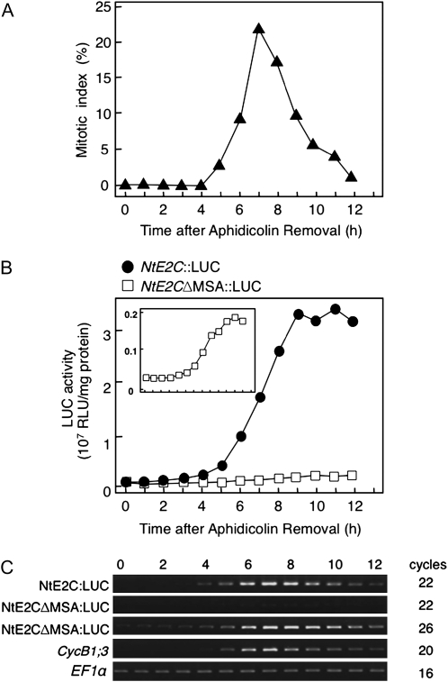
Functional analysis of the MSA element in the upstream region of the NtE2C gene. The LUC reporter gene was fused to either the wild-type NtE2C promoter (NtE2C∷LUC) or its mutant form lacking the MSA elements (NtE2CΔMSA∷LUC). BY-2 cells were transformed with these constructs and synchronized by aphidicolin treatment. After release from the aphidicolin block, the cells were sampled at 1-h intervals during culture. A, Change in the mitotic index in the synchronized culture. Mitotic index of the NtE2C∷LUC cells is shown. The NtE2CΔMSA∷LUC cells showed a similar pattern (data not shown). B, Change in LUC enzyme activity in the synchronized cultures. Protein extracts were prepared from each sample, and LUC enzyme activities were determined. The inset shows the change in the LUC activities of NtE2CΔMSA∷LUC cells with an expanded scale. RLU, Relative light units. C, Semiquantitative RT-PCR analysis of LUC mRNA levels in the synchronized culture. Total RNA was extracted from each sample and used for the RT-PCR analysis of LUC, CYCB1;3, and EF1α mRNAs. The EF1α mRNA was analyzed as a control that shows constitutive expression during the cell cycle. After electrophoresis in the presence of ethidium bromide, the gels were photographed under UV light illumination. Numbers above the gel images show time in hours after aphidicolin removal, and those on the right indicate numbers of PCR amplification cycles.

