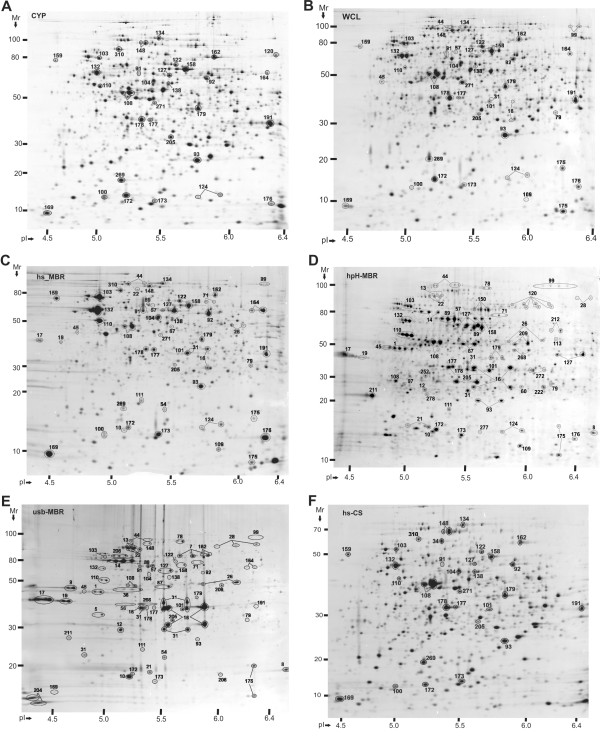
Figure 3.
A comparison of spot profiles in 2D gels derived from a Y. pestis KIM6+ whole cell lysate and five subcellular fractions. Acronyms are described in the flowchart of Figure 1. Cells were grown to stationary phase at 26°C. First dimension IEF separations were performed in the pH range from 4 to 7. The Mr range of second dimension SDS-PAGE separations was 10–200 kDa. Gel image analysis details are provided in the text. Spot identifications by MS confirmed appropriate spot matching. Spot numbers are equivalent to those denoted in Table 1; Additional File 2.

