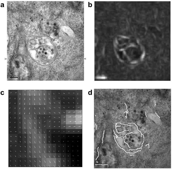
Figure 2.
Edge extraction on a microscopy image of a vesicle. An electron microscopy image of a vesicle (a), to which the edge extraction scheme is applied. The magnitude of the directional field extracted from the curvelet coefficients is shown in (b), with an extract showing the direction of the field in a small part of the image shown in (c). The last image (d) shows the final result of the edge extraction overlaid on the original image, after the edges have been extracted using the non-maximal suppression, and extended along the directional field to connect the different edge segments. Image courtesy of Prof. Urs Greber, University of Zürich.

