
Figure 1.
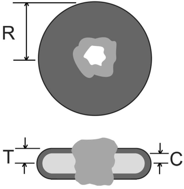
Schematic of the structure used for modeling a properly folded membrane protein in a membrane-like detergent micelle. The upper view shows the detergent disk (dark grey) of radius R penetrated by the structure of the membrane protein (grey) with a water channel through the structure (white). The lower figure shows a cut through the disk with the polar head group region of the detergent (dark grey) and nonpolar core of the bilayer (light grey), with thickness 2C. The micelle thickness is 2T.