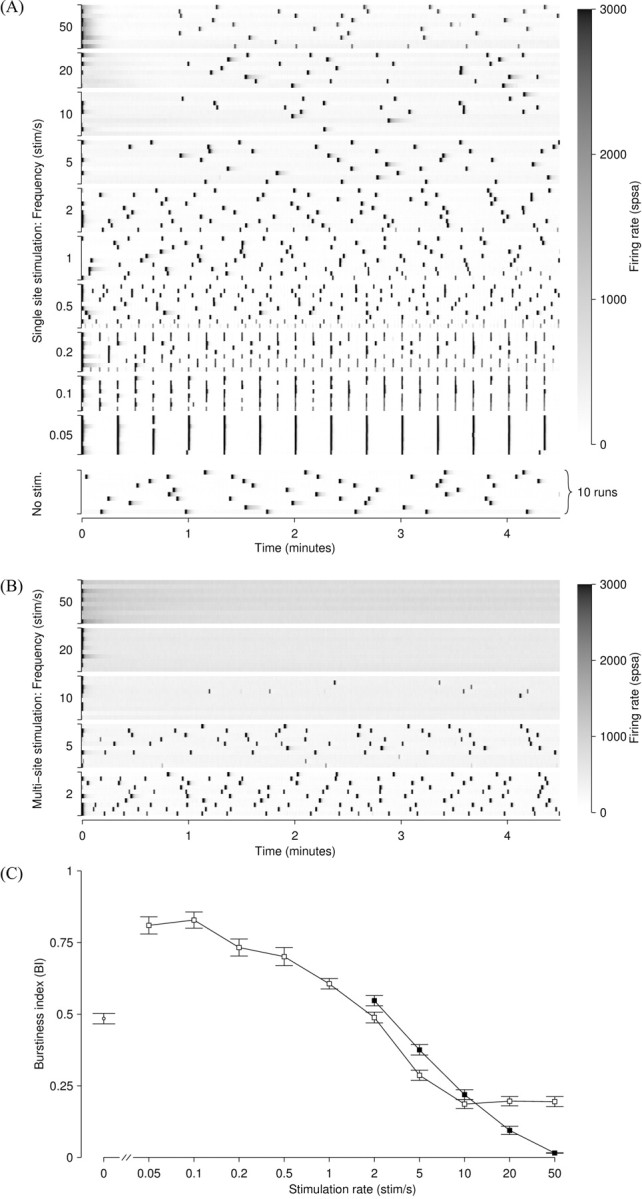
Figure 5.

A, Burstiness during single-electrode stimulation (protocolS) and spontaneous activity (no stimulation). Each row shows the array-wide firing rate (coded by the grayscale at right) as a function of time during one 5 min experimental run. In the 10 examples of spontaneous activity shown (bottom), bursts occurred irregularly approximately once per minute. In the 10 examples of stimulation at 0.05 stim/sec, bursts were perfectly aligned with stimuli, except in a few cases during which a spontaneous burst just preceded the stimulus. (The stimulating electrode was different in each of the 10 rows.) At 0.1-0.2 stim/sec, bursts underwent period doubling. Bursts during stimulation at 1-5 stim/sec were less frequent, but still mostly stimulus locked. In the 10-50 stim/sec runs, burst control was perfect for the first 45 sec, after which a spontaneous-like pattern returned. Data are from a culture at 39 DIV. Note that experimental runs were executed in random order. B, Bursting during multi-electrode stimulation (protocol M), same culture. Perfect and sustained burst control is attained at the higher stimulation frequencies. Note the increase in tonic firing rate (background shading) as the stimulation frequency is increased. C, Burstiness index as a function of stimulation frequency, for single-electrode stimulation (□) and multi-electrode stimulation (▪). Slow single-electrode stimulation elevates the burstiness over spontaneous (unstimulated) levels (○), whereas rapid stimulation reduces it. Values are mean ± SEM from n = 100 runs on 10 cultures. The most effective protocol tested, 50 stim/sec distributed across 25 electrodes, suppressed bursts completely (n = 60 runs, 6 cultures).
