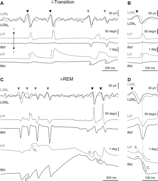
Figure 3.
Each part of the figure shows, from top to bottom, the activity in left and right lateral geniculate nucleus (LGNL and LGNR) (superimposed for easy comparison) and the velocity (LH', RH') and position (LH, RH) of the left and right eyes, respectively. Recording of the left and right LGN and eyes are in gray and black, respectively. A. During induced transition (i-Transition), primary ponto-geniculo-occipital waves (PGO) (head arrows) coincided with monocular rapid eye movements of the contralateral eye. C. During induced REM (i-REM) sleep, each cluster of complex-two-component (C1, C2) rapid eye movements coincided with a cluster of PGO waves. During i-REM, primary PGO waves occurred contralateral to the direction of the first component of the movement of the eye. B and D show an average (n = 40) of LGN activities and eye velocity and position triggered by the peak velocity of the isolated rapid eye movement during i-transition and by the second component of complex eye movement during i-REM sleep, respectively.

