
Fig. 2.
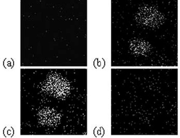
Positive ion molecular SIMS images of two separately dyed J774 cells following sputtering. (a) Composite image for DiD (m/z = 860, top) and DiI (m/z = 834, bottom). (b) Sims image for C5H9+ (m/z = 69). (c) SIMS image of PC head group ion (m/z = 184). (d) SIMS image of PE head group ion (m/z =142).