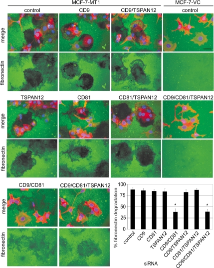
Figure 4.
Tetraspanin effects on MT1-MMP-dependent fibronectin degradation. MCF-7-VC and MCF-7-MT1 cells were treated with siRNAs, plated 4 d later on fibronectin (FN)-coated slides, and then incubated overnight in serum-free media with protease inhibitors (50 μg/ml aprotinin, 2 μM leupeptin, 20 μM E64, and 20 μM pepstatin A) to prevent non-MMP proteolysis of the FN. The following day, cells were washed, fixed, and FN was detected using an anti-FN antibody followed by Alexa 488-conjugated anti-mouse secondary antibody (green). The position of the cells was determined by staining the cytoskeleton with Alexa Fluor 546-phalloidin (red) and the nuclei with DAPI (blue). Representative fluorescent images (merge green/red/blue or green only) are shown. Bar, 20 μm. For the bar graph, 100% fibronectin degradation is defined as 0 pixel density within defined proteolyzed areas, whereas 0% degradation is pixel density in the unperturbed fibronectin layer. Results are mean of percentage of fibronectin degradation ± SD (n = 3; *p < 0.01 compared with control). Efficient tetraspanin knockdown was obtained similarly to that shown in Supplemental Figure 3.

