Figure 7.
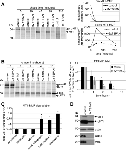
Tetraspanins affect MT1-MMP trafficking and stability. (A) MCF-7-MT1 cells were transfected with control or CD9/CD81/TSPAN12 (3xTSPAN) siRNAs. After 4 d, the cell surface subset of MT1-MMP was analyzed by [35S]Met/Cys pulse-chase (see Materials and Methods). Appearance and processing of cell surface MT1-MMP was quantitated by densitometry and plotted as raw densitometric values for both pro- and active MT1-MMP as line graphs. (B) Four days after siRNA treatment, total MT1-MMP was analyzed by [35S]Met/Cys pulse-chase, and densitometry was performed on total labeled MT1-MMP protein and expressed as ratio compared with control at time zero ± SD (n = 3; *p < 0.05 compared with control at each time point). cMT1 is cleaved MT1. (C) MCF-7-MT1-GFP cells were transfected with control or CD9/CD81/TSPAN12 (3xTSPAN) siRNAs. Two days after siRNA treatment, cells were treated with either lactacystin (10 μM), MG132 (2.5 μM), pepstatin A (50 μM)/leupeptin (10 μM), bafilomycin A1 (25 nM), or chloroquine (50 μM) for 2 d. Cell lysates were then collected, and MT1-MMP was analyzed by Western blotting. Densitometry was performed on total MT1-MMP and represented as ratio of total MT1-MMP for 3xTSPAN/control for each inhibitor ± SD (n = 3; *p < 0.05 compared with no inhibitor). Note that all the lysosomal inhibitors stimulated MT1-MMP expression in control siRNA-treated cells compared with no inhibitor, consistent with normal degradation of MT1-MMP in lysosomes (data not shown). (D) Tetraspanin knockdown was confirmed by Western blotting of CD9 and CD81 from cell lysates. Decreased MT1-MMP expression with tetraspanin knockdown is shown by Western blotting. TSPAN12 mRNA was also appropriately decreased (data not shown).

