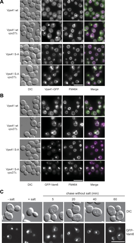
Figure 6.
Nonphosphorylated Vps41 and Vam6 are enriched at vacuole fusion sites. (A) Localization of Vps41-GFP in vps27Δ strain. SEY6210 cells expressing C-terminal GFP-tagged wild-type and mutant (S-A) Vps41 were labeled with FM4-64 and analyzed by fluorescence microscopy. Where indicated, VPS27 was deleted. Bar, 10 μm. (B) Localization of GFP-Vam6 in the vps27Δ strain. SEY6210 wild-type and vps27Δ cells expressing N-terminal GFP-tagged Vam6 in the respective Vps41 background were incubated with FM4-64 and analyzed by fluorescence microscopy. Bar, 10 μm. (C) In vivo fusion of vacuoles after relieve from high salt concentration stress. Cells expressing the Vps41 S-A mutant and the N-terminally GFP-tagged Vam6 fusion were incubated with 0.4 M NaCl for 30 min, followed by a wash with YPD medium and grown for the indicated times. Localization of Vam6 and vacuole morphology were monitored by fluorescence microscopy. Bar, 10 μm.

