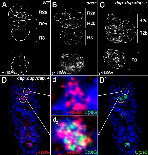
Figure 6.
dup/cdt1 dominantly enhances the DNA damage phenotype in dap−/− ovarian cysts. Wild-type (A), dap−/− mutant (B), and dap−, dupa1/dap−, + mutant germaria (C and D) stained with α-γ-H2Av (A–C; D, red) and C(3)G (D′, green) antibodies. The magnified images in the middle (d1 and d2) show C(3)G protein (green) and γ-H2Av foci (red) in one cell of a 16-cell cyst in early region 2a before the appearance of α-C(3)G staining (d1) and in one of the two pro-oocytes in region 2a after the appearance of α-C(3)G staining (d2). Note that γ-H2Av foci are increased in dap−, dupa1/dap−, + cysts (C and D) compared with wild-type (A) and dap−/− (B) cysts.

