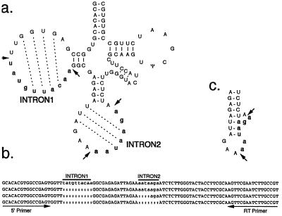
Figure 3.
Mapping introns of G. theta tRNASer gene. (a) Secondary structure of G. theta tRNASer showing introns 1 and 2 in bold lowercase. Intron splice sites are marked by arrows; dotted lines show potential base pairing between the intron and exon sequences. (b) Alignment of unspliced, partly spliced, and completely spliced reverse transcription–PCR products. Intron positions are indicated by black bars and the primers used for reverse transcription and PCR by black arrows. (c) Putative secondary structure of the extended anticodon stem and bulge structure containing the 3′ splice site.

