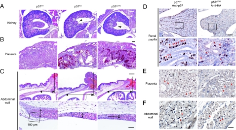
Fig. 5.
Tissue-specific residual phenotypes and corresponding expression patterns of knocked-in p27 in p57p27KI mice. (A and B) Representative histopathology of the renal papilla and placenta, respectively, of E18.5 embryos of the indicated genotypes. Hematoxylin–eosin staining shows developmental defects in the renal papilla (arrowheads), and necrosis in the placenta of p57KO and p57p27KI mice. (Scale bar, 200 μm.) (C) Representative histopathology of the umbilical region (Upper), and the tip of the rectus abdominis muscle (Lower) of P0 embryos. Arrows indicate the distance between the umbilicus and the tip of the rectus abdominis muscle (U-M distance) (Upper), and the thickness of the muscle layer at a position 100 μm from the tip (Lower). Hematoxylin–eosin staining reveals an increased U-M distance and thinner muscle layer in both p57KO and p57p27KI mice. Quantitative data are shown in Fig. S4. (Scale bars: for Upper, 200 μm; for Lower, 50 μm.) (D–F) Immunohistochemical analysis of the renal papilla (E18.5), placenta (E18.5), and abdominal wall (P0), respectively, of p57WT or p57p27KI mice performed with antibodies to p57 or to HA, respectively. Sections were counterstained with hematoxylin. Arrowheads indicate corresponding cells expressing either p57 or knocked-in p27. The boxed regions in D Upper are shown at higher magnification in the D Lower. T, spongiotrophoblast zone; L, labyrinth zone. (Scale bars: for D Upper, 100 μm; for D Lower, E, and F, 20 μm.)

