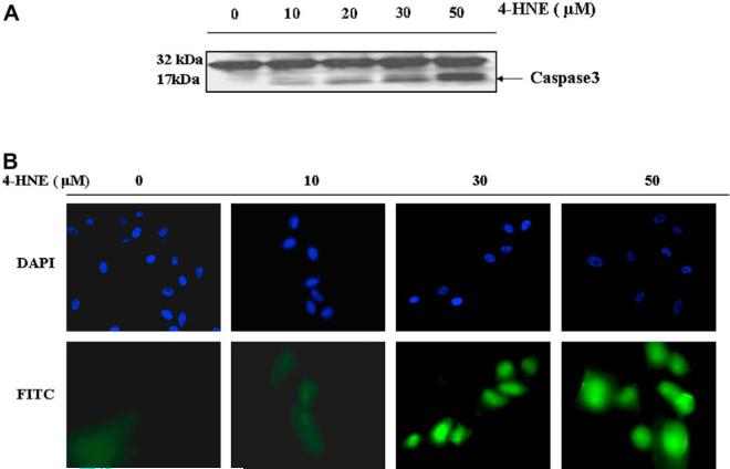
Fig. 5.
Effect of 4-HNE on caspase3 in RPE and ARPE-19 cells: (A) Cell extracts (50 μg protein) from RPE cells treated with 4-HNE (0-50 μM) for 2 h were resolved on 4-20% SDS-PAGE and immunoblotted using the anti-caspase3 antibody as the primary antibody. Activation of caspase3 was monitored by the appearance of the 20/17 kDa bands. The blot was developed using West Pico-chemiluminescence reagent (Pierce). (B) In situ analysis of activated caspase3 in ARPE-19 cells. About 2 × 10⁴ cells were grown in chamber slides and treated with 0, 10, 30, 50 μM 4-HNE for 2h. The activation of caspase3 in these cells was examined by staining with 10 μM CaspACE™ FITC-VAD-FMKin situ marker following the manufacturer's instructions. The slides were mounted with Vectashield DAPI mounting medium and observed with a fluorescence microscope (Olympus) using the standard filter sets for DAPI and FITC. Appropriately marked different panels show blue DAPI-stained and green FITC-stained cells in the figure. The photographs were taken at 400× magnification. (For interpretation of color mentioned in this figure the reader is referred to the web version of the article.)

