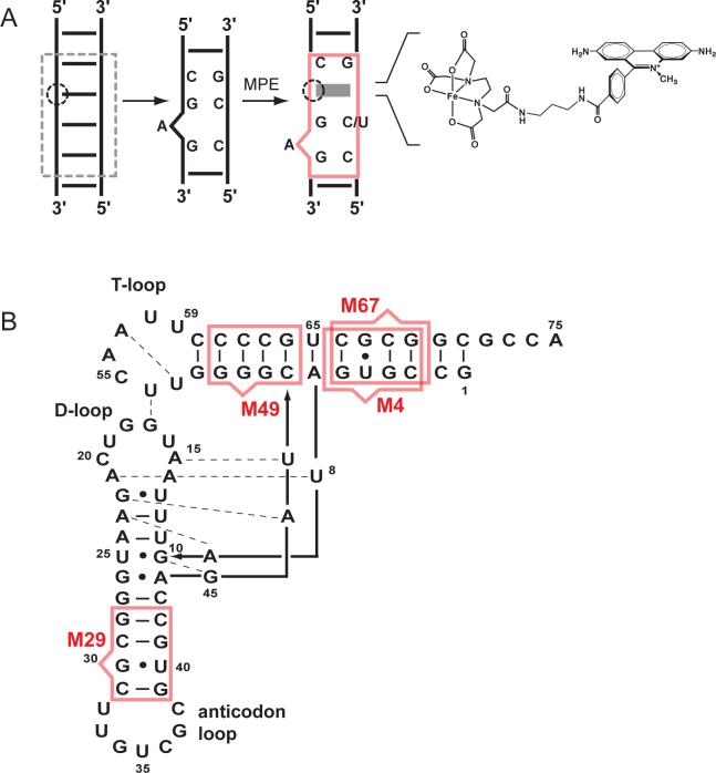
Figure 1. Analysis of RNA tertiary structure using a sequence-encoded cleavage agent.
(A). Intercalation of MPE in a bulged, three base-pair helix to replace four canonical base pairs (gray dashed square) in a helix (red square). MPE preferentially intercalates (solid gray box) such that the Fe(II)-EDTA moiety (dashed circle) is oriented towards the bulged A nucleotide. (B). tRNAAsp secondary structure and MPE binding sites. MPE binding sites are shown using the scheme from panel A. Mutants are numbered by the site nearest to the tethered Fe(II)-EDTA group.

