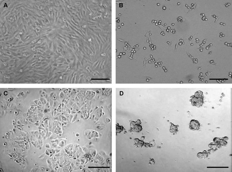
Fig. 2.

Photomicrographs showing cellular morphologies and growth properties of two tumor cell lines after 48 h of culture in the presence and absence of ECM. (A) A375.S2 (malignant melanoma), untreated; (B) A375.S2, co-cultured with ECM; (C) MCF7 (breast carcinoma), untreated; (D) MCF7, co-cultured with ECM. A and B, bar = 50 µm; C and D, bar = 100 µm.
