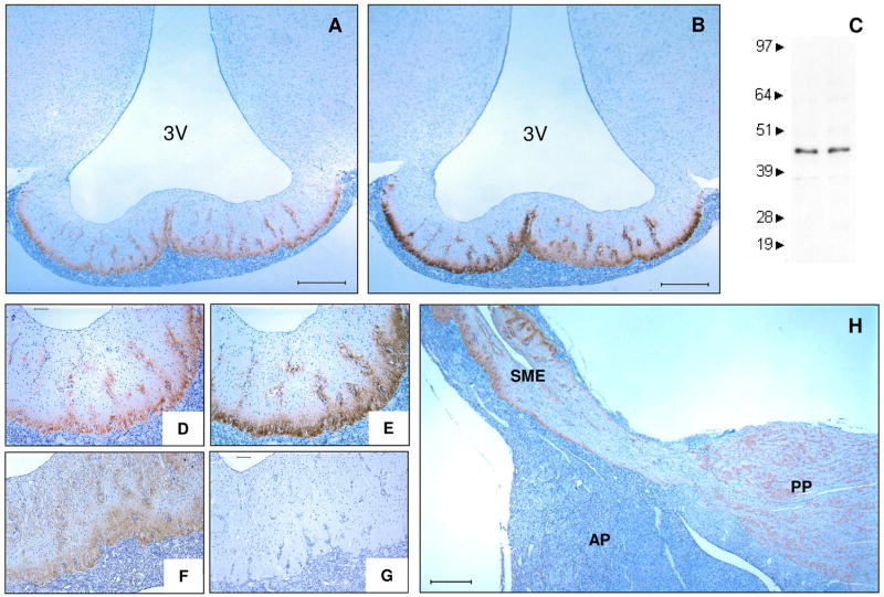
Figure 1.
Immunohistochemical detection of IL-6 in the external zone of the SME. Frontal view of IL-6 (A, D), CRH (B, E), VP (F) and non-immune control (G) in the SME (representative images, n ≥ 5 animals). Immunoblot analysis of IL-6 protein isolates from SME displaying immunoreactive bands at 45kDa (C). Sagittal view of pituitary showing IL-6 immunoreactivity in the external zone of the SME and the posterior pituitary (H). 3V, third ventricle; AP, anterior pituitary; PP, posterior pituitary; SME, stalk median eminence. Bar represents 500μm (A, B, H) and 100μm (D–G).

