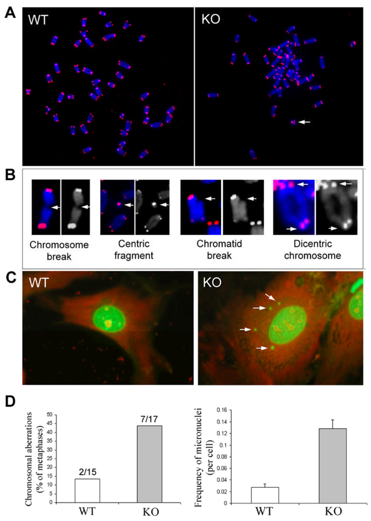
Figure 3. Increased frequency of chromosomal aberrations and micronuclei in early passage (P2) of TGFBI−/− MEFs.
(A) Digital images of Cy-3 (identify telomeres) and DAPI (identify chromosomes)-stained chromosomal metaphases in wild type and TGFBI KO MEFs. Arrow: centric ring. (B) Various types of chromosomal aberrations (Arrows) found in KO MEFs. (C) Multiple micronuclei (Arrows) identified in KO MEFs. (D) Frequency of chromosomal aberrations and micronuclei in wild type and TGFBI−/− MEFs.

