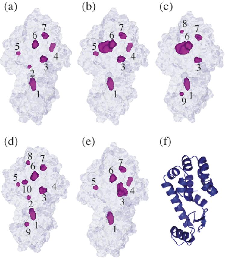
Figure 1.

T4 lysozyme structures shown from the same perspective. The external surface and buried cavities (shown in magenta) of (a) WT* (1L63), (b) L99A (1L90), (c) L99G/E108V (1QUH), (d) A98L (1QS5), and (e) V149G (1G0P) identified with a 1.2 Å probe in MSMS (34) with all internal solvent molecules removed. Cavities are identified by numbers 1 - 10 (refer to Table 2). (f) Cartoon representation of WT* T4 lysozyme. The C-terminal lobe is on the top side.
