
Figure 1.
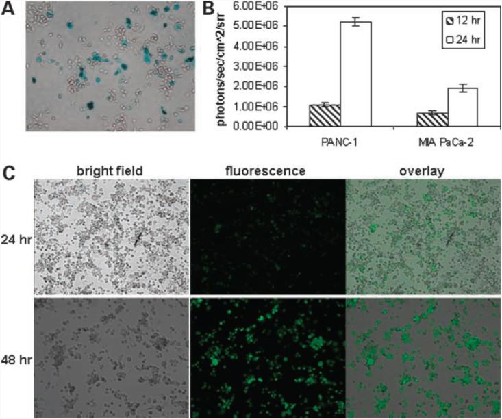
GLV-1h68 virus-mediated marker gene expressions in PANC-1 and MIA PaCa-2 cell cultures at different time points postinfection. A, β-gal staining (6 h) in PANC-1 cells. B, Renilla luciferase-catalyzed light emissions (12 and 24 h) in PANC-1 and MIA PaCa-2 cells. C, GFP expression (top, 24 h; bottom, 48 h) in PANC-1 cells.