
Figure 4.
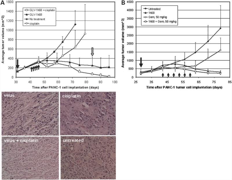
Combination therapy of s.c. PANC-1 tumors with GLV-1h68 virus and cisplatin or gemcitabine. A, combination of virus and cisplatin. Tumorous mice (n = 8 per group) were treated with the virus alone (1 × 10⁶ pfu), with cisplatin alone (6 mg/kg/d for 5 consecutive days), with the virus first (1 × 10⁶ pfu) followed by cisplatin treatment (6 mg/kg/d for 5 consecutive days) 10 d later, or with no treatment. Single solid arrow, time of virus injection; up-pointing arrows, time points of cisplatin treatments. For histology analysis, mice were killed 55 d postvirus injection. Tumors were excised, sectioned, and stained with H&E. Open arrow, time point of histologic analyses (n = 8 per group). Microscopic analyses of virus (top left), cisplatin (top right), and combination therapy (bottom left) treated and untreated (bottom right) tumor sections (5 μm) stained with H&E (bottom). Magnification, ×200. Also shown are examples of two mice after their s.c. PANC-1 tumors were eradicated by the combination therapy. B, combination of virus and gemcitabine. Single solid arrow, time of virus injection; up-pointing arrows, time points of gemcitabine treatments (n = 8 per group).