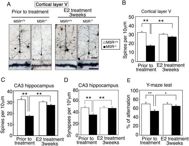
Figure 7. E2 restores normal morphology in cortical and hippocampal pyramidal neurons from male M5R−/− mice.
(A) Morphologic changes of cortical pyramidal neurons (layer V) from male M5R−/− mice without or after chronic E2 treatment (3 weeks). Golgi staining revealed that cortical pyramidal neurons from male M5R−/− mice showed clear signs of atrophy of the basal-dendritic tree and apical dendrites. E2 treated male M5R−/− mice showed a similar morphology of spines in the basal-dendritic tree and apical dendrites as M5R+/+ control mice. (B) Number of spines per 10 µm length of dendritic segment of cortical pyramidal neurons (layer V) from male M5R−/− and M5R+/+ mice, and E2 treated male M5R−/− and M5R+/+ mice. N = 40 dendritic segments from 5 animals per group. (C) Number of spines per 10 µm length of dendritic segment of CA3 hippocampal pyramidal neurons from male M5R−/− and M5R+/+ mice, and E2 treated male M5R−/− and M5R+/+ mice. Hippocampal neurons from male M5R−/− mice exhibited a significantly reduced number of dendritic spines. E2 administration also restored the number of dendritic spines in the CA3 hippocampus. N = 20 dendritic segments from 5 animals per group. Values are means±SEM. **p<0.001. (D) The number of synapses per 102 µm was counted in the CA3 hippocampus by analyzing electron microscope images. A total of 100–110 sections of 70 nm electron microscope images were counted for each sample. Values are means±SEM. **p<0.001. (E) Performance of male M5R+/+ and M5R−/− mice prior to and after chronic E2 treatment in a Y-maze spatial-memory test. Chronic E2 tablet (0.1 mg / 21 days release) treatment completely rescues cerebrovascular and cognitive deficits in male M5R−/− mice. All studies were carried out with 3 month-old male M5R+/+ (n = 16) and M5R−/− (n = 14) mice. Values are means±SEM. *p<0.05, **p<0.001.

