
FIGURE 2. Histopathological examination of full-thickness incisional wounds at days 3, 7 and 14 after surgery and treatment.
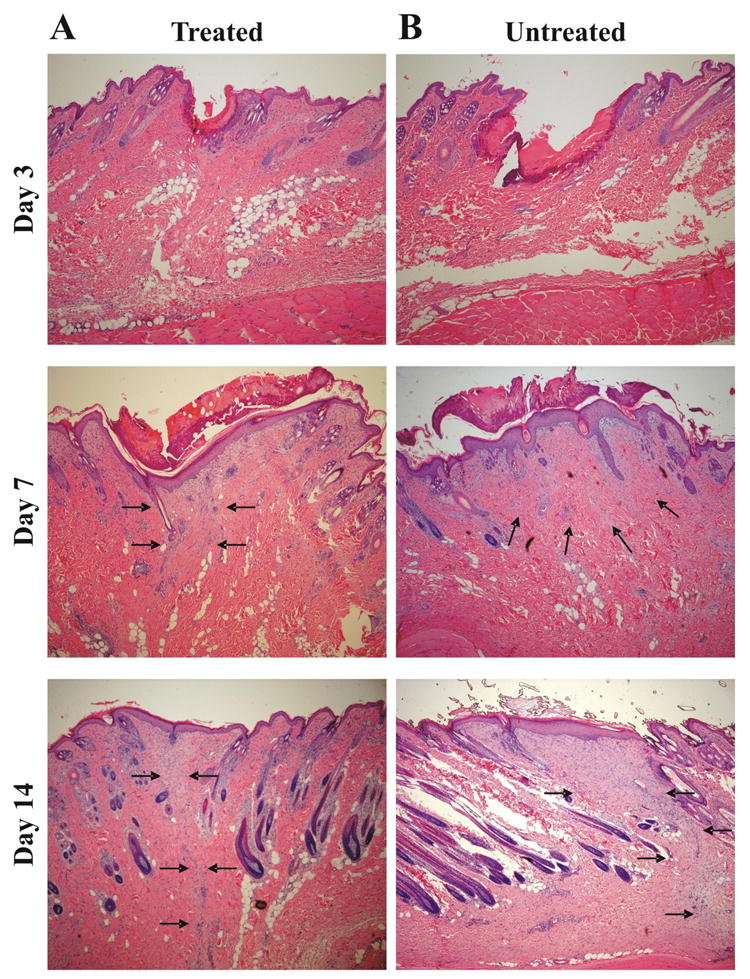
Histopathology of the (A) hMSC treated group and (B) control group at 3, 7 and 14 days after the operation (H&E stain, 40×). Tissue sections were stained with hematoxylin and eosin and examined for the differences in epithelial gap and granulation tissue by a Pathologist blinded to the treatment groups. No differences in epithelial gap could be distinguished among the three treatment groups at day 3 and 7 after surgery. At day 14 after surgery and treatment the granulation tissue area was smaller in the hMSC treated wounds (A) compared with untreated wounds (B). In none of the groups signs of inflammation could be detected. Arrows indicate to granulation tissue.