
FIGURE 3. Histopathological examination of full-thickness incisional wounds at days 21and 80 after surgery and treatment.
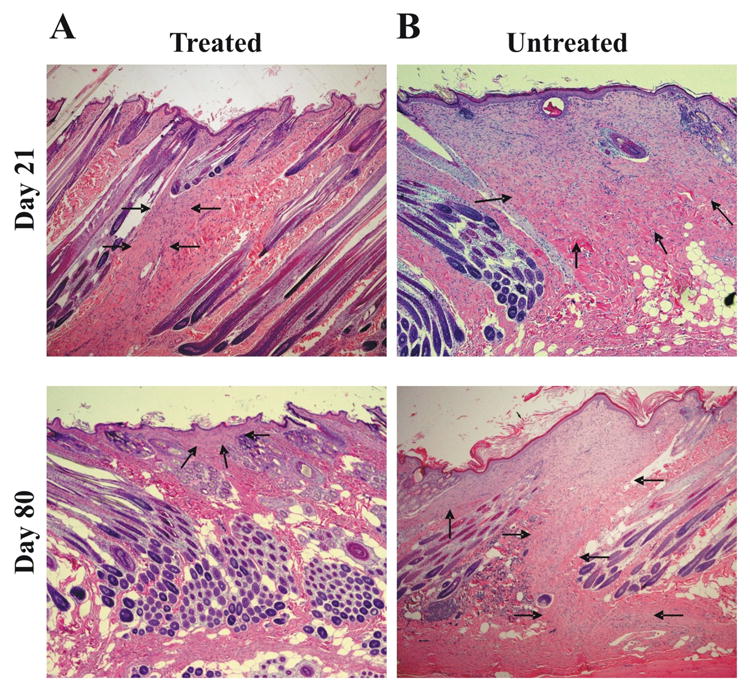
Histopathology of the (A) hMSC treated group and (B) control group at 21 and 80 days after the operation (H&E stain, 40×). Tissue sections were stained with hematoxylin and eosin and examined for the granulation tissue by a pathologist blinded to the treatment. The width of the granulation tissue is narrower in the hMSC treated group (A) than the control group (B) at days 21 and 80 after surgery. In none of the groups signs of inflammation could be detected. Arrows indicate to granulation tissue.