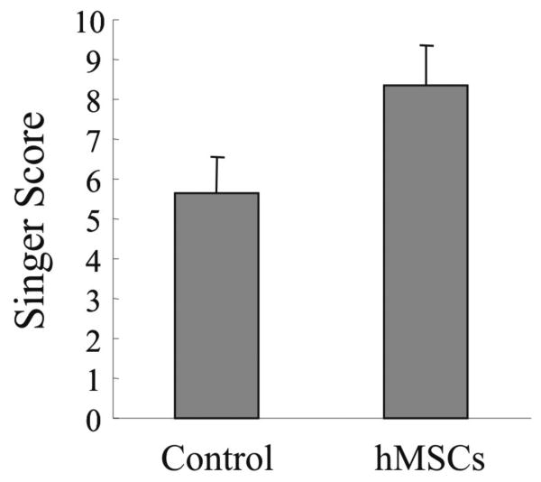
FIGURE 4. Histomorphologic evaluation of the incisional wounds by Singer classification at day 80 after surgery and treatment.
Incisional wounds treated with hMSCs or untreated wounds (control) were evaluated for the presence of hyperkeratosis, epidermal hyperplasia, presence and depth of collagen disorganization, fibroplasia, vascular proliferation, absence of adnexa, including hair follicles, apocrine glands, and smooth muscle according to Singer [14] at day 80 after treatment. The scores ranged from 0 (worst scarring) to 10 (absence of scarring). Results represent the mean ± SD; n = 3 rabbits at day 80. * p < 0.05 hMSC treated group versus control group.

