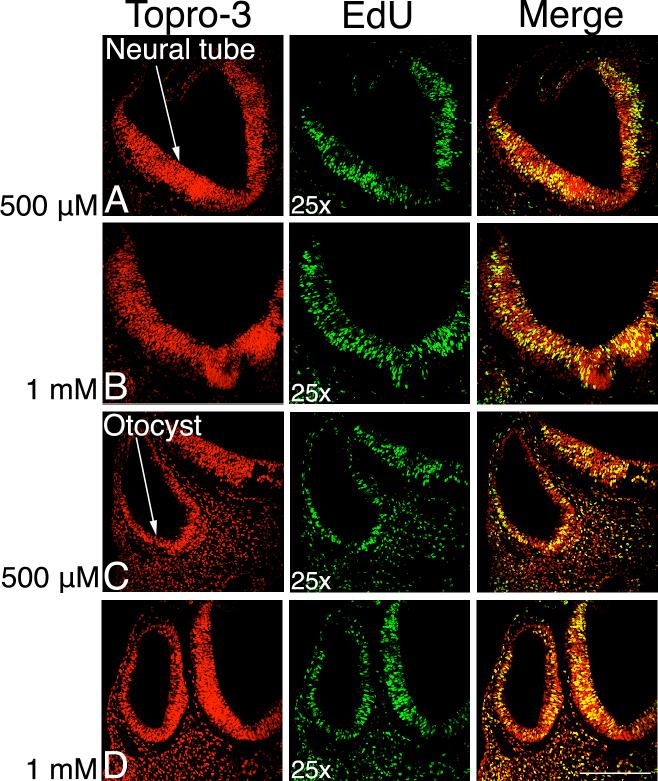
Figure 1. EdU labels proliferating cells in day 3 chick embryonic tissue.
Dorsal to the top, 10 μm paraffin sections labeled with TO-PRO-3, a DNA marker labeling every cell, in red. EdU is in green, with the merged images in the right hand column. The yellow color in the merged image shows the proliferating cells. Rows A and C labeled with 500 μM EdU, B and D with 1 mM. The neural tube (nt) and otocyst (o) are labeled. Scale bar: 200 μm.

