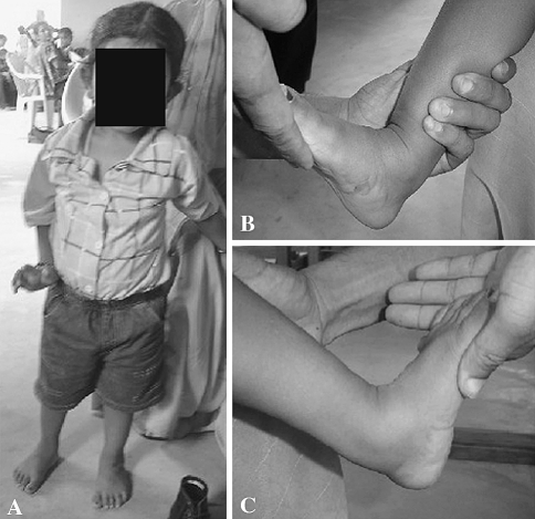
Fig. 3A–C.
A patient treated by the Ponseti method for bilateral idiopathic clubfoot is shown with followup photograph (A) standing and (B–C) with maximal passive dorsiflexion. (A) This boy has just completed the Ponseti method for a bilateral clubfoot deformity and has achieved a heel-toe gait. (B) Maximum passive dorsiflexion on the right side. (C) Maximum passive dorsiflexion on the left side.

