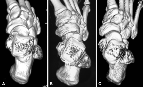
Fig. 1A–C.
Three-dimensional computed tomography scan reconstruction of the foot in the transverse plane at the level of the ankle mortise in (A) a normal foot, (B) a clubfoot of the first series, and (C) a clubfoot of the second series. The torsion angle of the ankle measured 19° in the normal foot, 30.5° in the clubfoot of the first series, and 26.5° in the clubfoot of the second series. The declination angle of the neck of the talus measured 18° in the normal foot, 31° in the first series clubfoot, and 23.5 in the second series clubfoot. A residual varus deformity of the calcaneus is present in the clubfoot of the first series. The navicular tuberosity is very close to the medial malleolus in the clubfoot of the second series, but the cuneiforms and the cuboid are shifted laterally.

