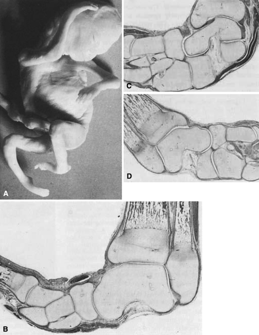
Fig. 1A–D.
(A) 90-mm (crown to rump) male fetus with bilateral clubfoot. (B) Frontal section of the left foot showing the talus in the ankle mortise, and the navicular, cuneiforms, and first and second metatarsals displaced medially. The anterior tibial tendon is seen above the navicular. Endochondral ossification is seen in the tibia, fibula and first metatarsal (×15). (C) Frontal section of the left foot taken about 3 mm behind section (B). The anterior and posterior portions of the os calcis are underneath the talus. The navicular articulates with the medial aspect of the head of the talus. The three cuneiforms are to the left and the cuboid is underneath the navicular. The medial ankle ligament is folded (×15). (D) Frontal section of the left foot. The talus is in the ankle mortise. The navicular is medial and the anterior portion of the os calcis is underneath the head of the talus. The deltoid ligament is slightly folded following the contour of the head of the talus. A branch of the posterior tibial tendon is seen directed toward the base of the fourth metatarsal. In (C) and (D) there is a cellular connection between the calcaneus and the navicular suggesting a calcaneonavicular fusion (×15).

